• 文献检索
  • 文档翻译
  • 深度研究
  • 学术资讯
  • Suppr Zotero 插件Zotero 插件
  • 邀请有礼
  • 套餐&价格
  • 历史记录
应用&插件
Suppr Zotero 插件Zotero 插件浏览器插件Mac 客户端Windows 客户端微信小程序
定价
高级版会员购买积分包购买API积分包
服务
文献检索文档翻译深度研究API 文档MCP 服务
关于我们
关于 Suppr公司介绍联系我们用户协议隐私条款
关注我们

Suppr 超能文献

核心技术专利:CN118964589B侵权必究
粤ICP备2023148730 号-1Suppr @ 2026

文献检索

告别复杂PubMed语法,用中文像聊天一样搜索,搜遍4000万医学文献。AI智能推荐,让科研检索更轻松。

立即免费搜索

文件翻译

保留排版,准确专业,支持PDF/Word/PPT等文件格式,支持 12+语言互译。

免费翻译文档

深度研究

AI帮你快速写综述,25分钟生成高质量综述,智能提取关键信息,辅助科研写作。

立即免费体验

白化小鼠光损伤后的视网膜代偿性变化。

Retinal compensatory changes after light damage in albino mice.

作者信息

Montalbán-Soler Luis, Alarcón-Martínez Luis, Jiménez-López Manuel, Salinas-Navarro Manuel, Galindo-Romero Caridad, Bezerra de Sá Fabrízio, García-Ayuso Diego, Avilés-Trigueros Marcelino, Vidal-Sanz Manuel, Agudo-Barriuso Marta, Villegas-Pérez Maria P

机构信息

Departamento de Oftalmología, Optometría, Otorrinolaringología y Anatomía Patológica, Facultad de Medicina, Universidad de Murcia, Murcia, Spain.

出版信息

Mol Vis. 2012;18:675-93. Epub 2012 Mar 24.

PMID:22509098
原文链接:https://pmc.ncbi.nlm.nih.gov/articles/PMC3325904/
Abstract

PURPOSE

To investigate the anatomic and functional changes triggered by light exposure in the albino mouse retina and compare them with those observed in the albino rat.

METHODS

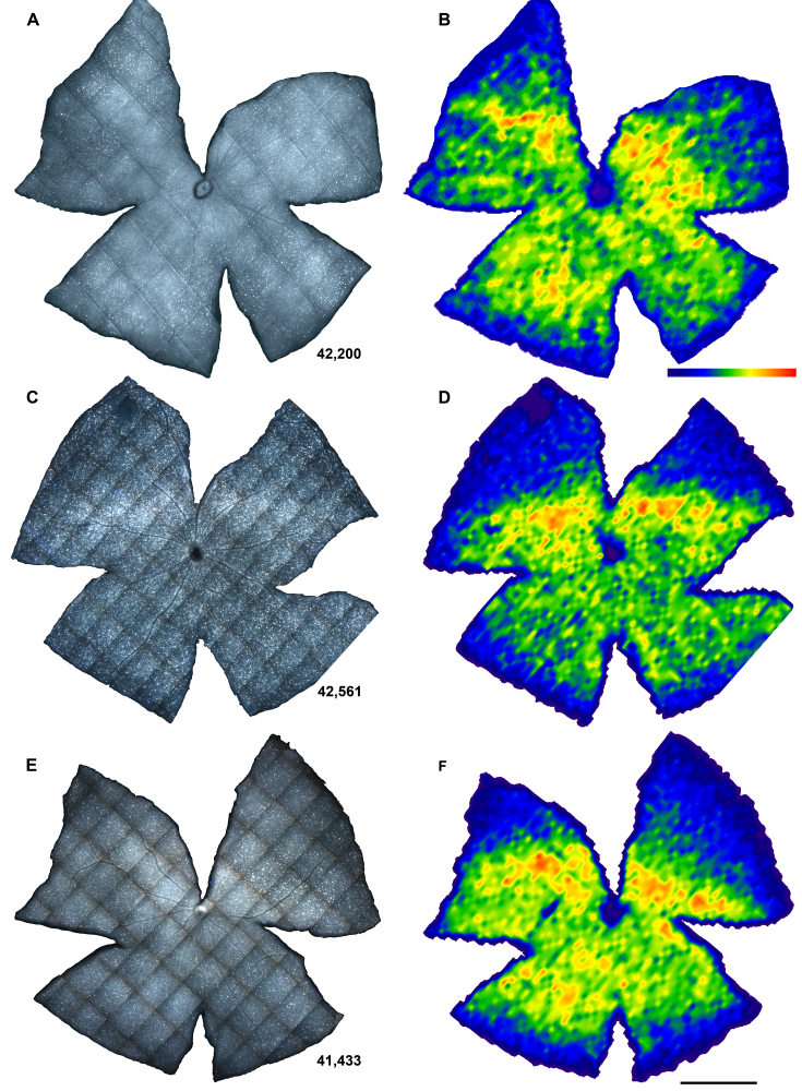
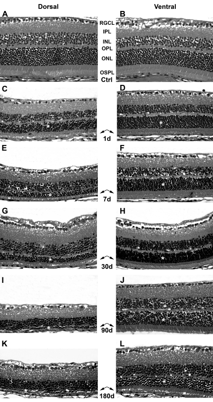
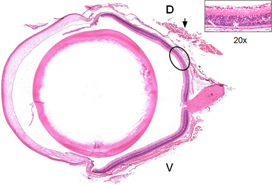
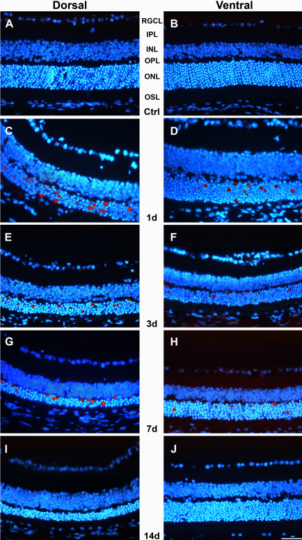
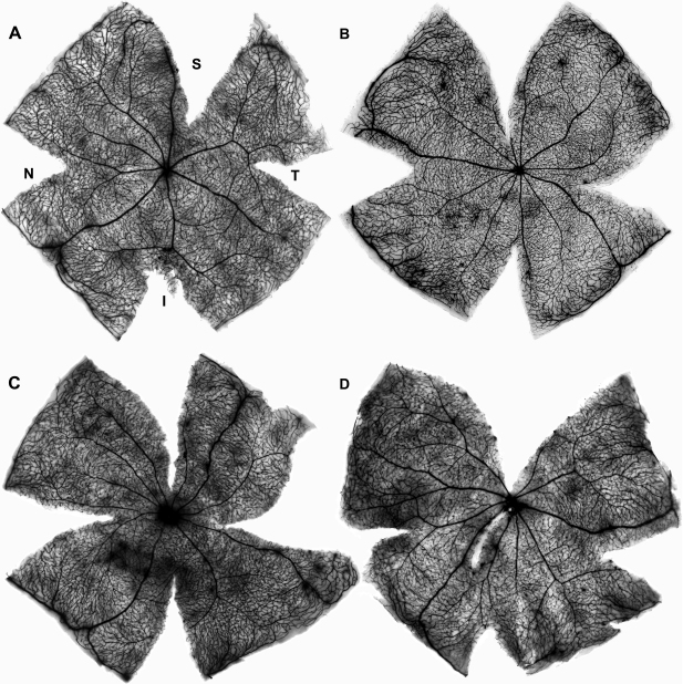
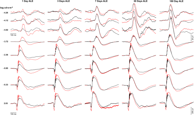
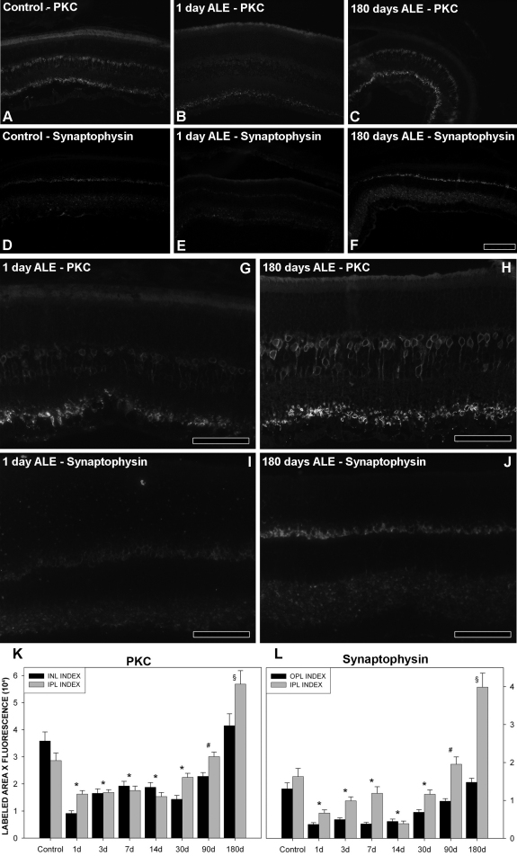
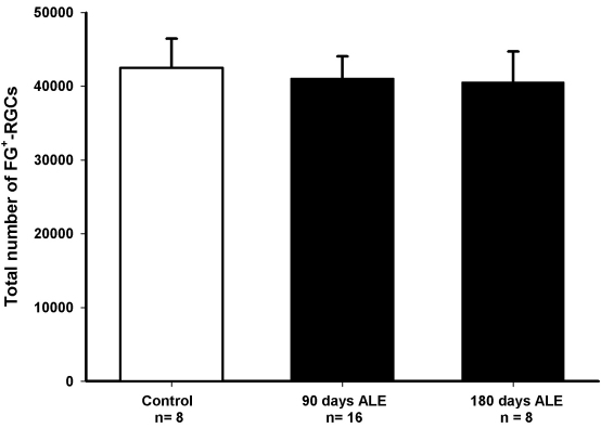
BALB/c albino mice were exposed to 3,000 lx of white light during 24 h and their retinas analyzed from 1 to 180 days after light exposure (ALE). Left pupil mydriasis was induced with topical atropine. Retinal function was analyzed by electroretinographic (ERG) recording. To assess retinal degeneration, hematoxylin and eosin staining, the TdT-mediated dUTP nick-end labeling (TUNEL) technique, and quantitative immunohistofluorescence for synaptophysin and protein kinase Cα (PKCα) were used in cross sections. Intravenous injection of horseradish peroxidase and Fluoro-Gold™ tracing were used in whole-mounted retinas to study the retinal vasculature and the retinal ganglion cell (RGC) population, respectively.

RESULTS

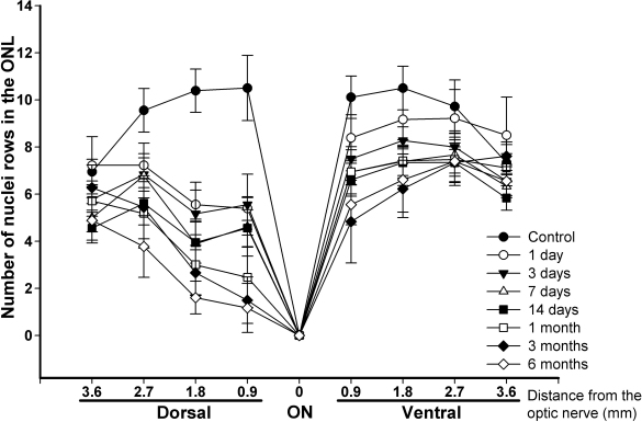
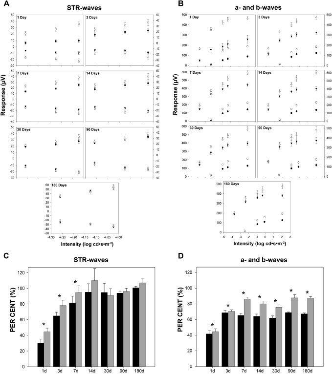
Light exposure caused apoptotic photoreceptor death in the central retina. This death was more severe in the dorsal than in the ventral retina, sparing the periphery. Neither retinal vascular leakage nor retinal ganglion cell death was observed ALE. The electroretinographic a-wave was permanently impaired, while the b-wave decreased but recovered gradually by 180 days ALE. The scotopic threshold responses, associated with the inner retinal function, diminished at first but recovered completely by 14 days ALE. This functional recovery was concomitant with the upregulation of protein kinase Cα and synaptophysin. Similar results were obtained in both eyes, irrespective of mydriasis.

CONCLUSIONS

In albino mice, light exposure induces substantial retinal damage, but the surviving photoreceptors, together with compensatory morphological/molecular changes, allow an important restoration of the retinal function.

摘要

目的

研究光照对白化病小鼠视网膜引发的解剖学和功能变化,并将其与白化病大鼠中观察到的变化进行比较。

方法

将BALB/c白化病小鼠暴露于3000勒克斯的白光下24小时,并在光照后1至180天(ALE)对其视网膜进行分析。用局部阿托品诱导左瞳孔散大。通过视网膜电图(ERG)记录分析视网膜功能。为评估视网膜变性,在横切面上使用苏木精和伊红染色、TdT介导的dUTP缺口末端标记(TUNEL)技术以及对突触素和蛋白激酶Cα(PKCα)的定量免疫荧光。在整个视网膜标本中分别静脉注射辣根过氧化物酶和Fluoro-Gold™示踪剂,以研究视网膜血管系统和视网膜神经节细胞(RGC)群体。

结果

光照导致中央视网膜中的光感受器凋亡性死亡。这种死亡在背侧视网膜比腹侧视网膜更严重,周边区域未受影响。在ALE时未观察到视网膜血管渗漏或视网膜神经节细胞死亡。视网膜电图的a波永久性受损,而b波降低,但在ALE后180天逐渐恢复。与视网膜内层功能相关的暗视阈值反应起初降低,但在ALE后14天完全恢复。这种功能恢复与蛋白激酶Cα和突触素的上调同时发生。无论瞳孔散大与否,双眼均获得相似结果。

结论

在白化病小鼠中,光照会引起显著的视网膜损伤,但存活的光感受器以及代偿性的形态学/分子变化使得视网膜功能得到重要恢复。

https://cdn.ncbi.nlm.nih.gov/pmc/blobs/6c08/3325904/42d5d45fa27c/mv-v18-675-f10.jpg
https://cdn.ncbi.nlm.nih.gov/pmc/blobs/6c08/3325904/943edd7acb8a/mv-v18-675-f1.jpg
https://cdn.ncbi.nlm.nih.gov/pmc/blobs/6c08/3325904/d35f76c42f34/mv-v18-675-f2.jpg
https://cdn.ncbi.nlm.nih.gov/pmc/blobs/6c08/3325904/5affe1133d11/mv-v18-675-f3.jpg
https://cdn.ncbi.nlm.nih.gov/pmc/blobs/6c08/3325904/3b19a291d6bf/mv-v18-675-f4.jpg
https://cdn.ncbi.nlm.nih.gov/pmc/blobs/6c08/3325904/496d7adb9367/mv-v18-675-f5.jpg
https://cdn.ncbi.nlm.nih.gov/pmc/blobs/6c08/3325904/ea3dae784c9b/mv-v18-675-f6.jpg
https://cdn.ncbi.nlm.nih.gov/pmc/blobs/6c08/3325904/c30eb753089b/mv-v18-675-f7.jpg
https://cdn.ncbi.nlm.nih.gov/pmc/blobs/6c08/3325904/a8ba18a9d967/mv-v18-675-f8.jpg
https://cdn.ncbi.nlm.nih.gov/pmc/blobs/6c08/3325904/596578937a76/mv-v18-675-f9.jpg
https://cdn.ncbi.nlm.nih.gov/pmc/blobs/6c08/3325904/42d5d45fa27c/mv-v18-675-f10.jpg

相似文献

1
Retinal compensatory changes after light damage in albino mice.白化小鼠光损伤后的视网膜代偿性变化。
Mol Vis. 2012;18:675-93. Epub 2012 Mar 24.
2
Retinal ganglion cell axonal compression by retinal vessels in light-induced retinal degeneration.光诱导性视网膜变性中视网膜血管对视网膜神经节细胞轴突的压迫
Mol Vis. 2011;17:1716-33. Epub 2011 Jun 25.
3
Quantitative relationship of the scotopic and photopic ERG to photoreceptor cell loss in light damaged rats.暗视和明视视网膜电图与光损伤大鼠光感受器细胞丢失的定量关系。
Exp Eye Res. 2000 May;70(5):693-705. doi: 10.1006/exer.2000.0842.
4
Light-induced cell death of retinal photoreceptors in the absence of p53.在缺乏p53的情况下,光诱导的视网膜光感受器细胞死亡。
Invest Ophthalmol Vis Sci. 1998 Apr;39(5):846-9.
5
Functional and morphological evaluation of blue light-emitting diode-induced retinal degeneration in mice.蓝光发光二极管诱导的小鼠视网膜变性的功能和形态学评估
Graefes Arch Clin Exp Ophthalmol. 2016 Apr;254(4):705-16. doi: 10.1007/s00417-015-3258-x. Epub 2016 Jan 8.
6
Light-induced retinal degeneration causes a transient downregulation of melanopsin in the rat retina.光诱导的视网膜变性导致大鼠视网膜中黑视蛋白的短暂下调。
Exp Eye Res. 2017 Aug;161:10-16. doi: 10.1016/j.exer.2017.05.010. Epub 2017 May 26.
7
Suppression of developmental retinal cell death but not of photoreceptor degeneration in Bax-deficient mice.Bax基因缺陷小鼠中发育性视网膜细胞死亡受到抑制,但光感受器退化未受抑制。
Invest Ophthalmol Vis Sci. 1998 Aug;39(9):1713-20.
8
Berberine protects against light-induced photoreceptor degeneration in the mouse retina.黄连素可保护小鼠视网膜免受光诱导的光感受器退化。
Exp Eye Res. 2016 Apr;145:1-9. doi: 10.1016/j.exer.2015.10.005. Epub 2015 Oct 22.
9
Increased sensitivity to light-induced damage in a mouse model of autosomal dominant retinal disease.常染色体显性视网膜疾病小鼠模型中对光诱导损伤的敏感性增加。
Invest Ophthalmol Vis Sci. 2007 May;48(5):1942-51. doi: 10.1167/iovs.06-1131.
10
Systemic administration of the antioxidant/iron chelator α-lipoic acid protects against light-induced photoreceptor degeneration in the mouse retina.抗氧化剂/铁螯合剂α-硫辛酸的全身给药可保护小鼠视网膜免受光诱导的光感受器退化。
Invest Ophthalmol Vis Sci. 2014 Aug 21;55(9):5979-88. doi: 10.1167/iovs.14-15025.

引用本文的文献

1
Oral 8-aminoguanine against age-related retinal degeneration.口服8-氨基鸟嘌呤治疗年龄相关性视网膜变性。
Commun Biol. 2025 May 26;8(1):812. doi: 10.1038/s42003-025-08242-1.
2
Accessible LED Lightbox for Light-induced Retinal Damage in Pigmented Mice.用于诱导色素沉着小鼠视网膜损伤的简易LED灯箱
Invest Ophthalmol Vis Sci. 2025 Feb 3;66(2):49. doi: 10.1167/iovs.66.2.49.
3
Oral 8-aminoguanine against age-related retinal degeneration.口服8-氨基鸟嘌呤治疗年龄相关性视网膜变性。

本文引用的文献

1
Retinal ganglion cell axonal compression by retinal vessels in light-induced retinal degeneration.光诱导性视网膜变性中视网膜血管对视网膜神经节细胞轴突的压迫
Mol Vis. 2011;17:1716-33. Epub 2011 Jun 25.
2
Axotomy-induced retinal ganglion cell death in adult mice: quantitative and topographic time course analyses.成年小鼠视神经切断诱导的视网膜神经节细胞死亡:定量和地形时程分析。
Exp Eye Res. 2011 May;92(5):377-87. doi: 10.1016/j.exer.2011.02.008. Epub 2011 Feb 24.
3
Retinal ganglion cell numbers and delayed retinal ganglion cell death in the P23H rat retina.
Res Sq. 2024 May 6:rs.3.rs-4022389. doi: 10.21203/rs.3.rs-4022389/v1.
4
Comparison between sodium iodate and lipid peroxide murine models of age-related macular degeneration for drug evaluation-a narrative review.用于药物评估的碘酸钠和脂质过氧化小鼠年龄相关性黄斑变性模型的比较——一篇叙述性综述
Ann Eye Sci. 2022 Mar 15;7. doi: 10.21037/aes-21-25.
5
Cellular and Molecular Mechanisms of Pathogenesis Underlying Inherited Retinal Dystrophies.遗传性视网膜疾病发病机制的细胞和分子机制。
Biomolecules. 2023 Feb 1;13(2):271. doi: 10.3390/biom13020271.
6
Analysis of mitochondrial dynamics and function in the retinal pigment epithelium by high-speed high-resolution live imaging.通过高速高分辨率实时成像分析视网膜色素上皮细胞中的线粒体动力学和功能。
Front Cell Dev Biol. 2022 Oct 28;10:1044672. doi: 10.3389/fcell.2022.1044672. eCollection 2022.
7
Neural and Müller glial adaptation of the retina to photoreceptor degeneration.视网膜对光感受器变性的神经和米勒胶质细胞适应性变化
Neural Regen Res. 2023 Apr;18(4):701-707. doi: 10.4103/1673-5374.354511.
8
ERG and Behavioral CFF in Light-Damaged Albino Rats.ERG 和行为性 CFF 在光损伤白化大鼠中的变化。
Int J Mol Sci. 2022 Apr 8;23(8):4127. doi: 10.3390/ijms23084127.
9
Cone Photoreceptor Loss in Light-Damaged Albino Rats.光损伤白化大鼠的视锥光感受器损失
Int J Mol Sci. 2022 Apr 2;23(7):3978. doi: 10.3390/ijms23073978.
10
Animal Models of LED-Induced Phototoxicity. Short- and Long-Term In Vivo and Ex Vivo Retinal Alterations.LED诱导的光毒性动物模型。短期和长期体内及体外视网膜改变。
Life (Basel). 2021 Oct 26;11(11):1137. doi: 10.3390/life11111137.
P23H 大鼠视网膜中的神经节细胞数量和神经节细胞延迟死亡。
Exp Eye Res. 2010 Dec;91(6):800-10. doi: 10.1016/j.exer.2010.10.003. Epub 2010 Oct 16.
4
ERG changes in albino and pigmented mice after optic nerve transection.视神经横断后白化病小鼠和有色小鼠的视网膜电图变化。
Vision Res. 2010 Oct 12;50(21):2176-87. doi: 10.1016/j.visres.2010.08.014. Epub 2010 Aug 19.
5
Changes in the inner and outer retinal layers after acute increase of the intraocular pressure in adult albino Swiss mice.成年白化瑞士小鼠眼内压急性升高后内外视网膜层的变化。
Exp Eye Res. 2010 Aug;91(2):273-85. doi: 10.1016/j.exer.2010.05.020. Epub 2010 Jun 1.
6
Photoreceptor degeneration: genetic and mechanistic dissection of a complex trait.光感受器变性:复杂性状的遗传与机制剖析。
Nat Rev Genet. 2010 Apr;11(4):273-84. doi: 10.1038/nrg2717.
7
Late histological and functional changes in the P23H rat retina after photoreceptor loss.P23H 大鼠视网膜感光细胞丧失后晚期的组织学和功能变化。
Neurobiol Dis. 2010 Apr;38(1):47-58. doi: 10.1016/j.nbd.2009.12.025. Epub 2010 Jan 7.
8
Retinal light damage: mechanisms and protection.视网膜光损伤:机制与保护。
Prog Retin Eye Res. 2010 Mar;29(2):113-34. doi: 10.1016/j.preteyeres.2009.11.004. Epub 2009 Dec 3.
9
Retinal ganglion cell population in adult albino and pigmented mice: a computerized analysis of the entire population and its spatial distribution.成年白化小鼠和有色小鼠的视网膜神经节细胞群体:对整个群体及其空间分布的计算机分析。
Vision Res. 2009 Mar;49(6):637-47. doi: 10.1016/j.visres.2009.01.010. Epub 2009 Jan 29.
10
Short and long term axotomy-induced ERG changes in albino and pigmented rats.白化和有色大鼠短期和长期轴突切断诱导的视网膜电图变化。
Mol Vis. 2009 Nov 17;15:2373-83.