Florian A, Jurcut R, Ginghina C, Bogaert J
Carol Davila University of Medicine and Pharmacy, Bucharest, Romania.
J Med Life. 2011 Nov 14;4(4):330-45. Epub 2011 Nov 24.
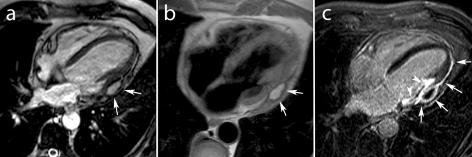
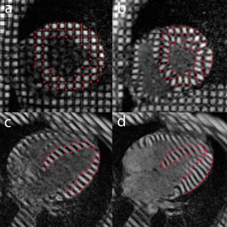
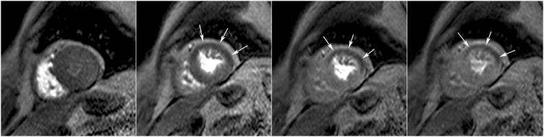
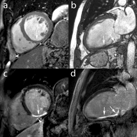
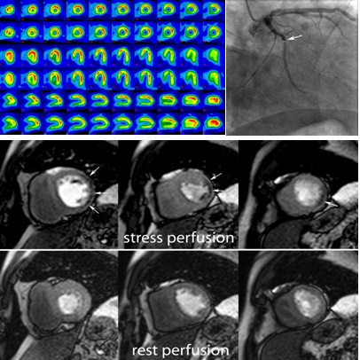
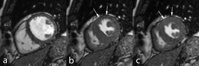
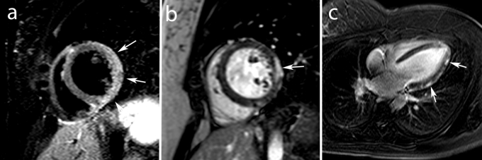
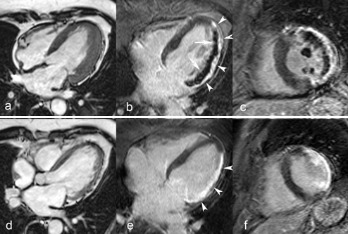
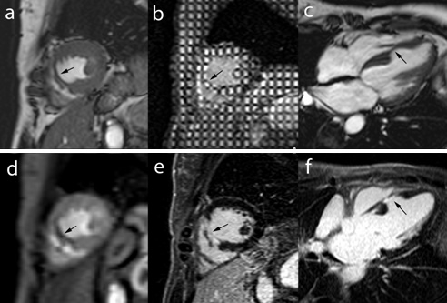
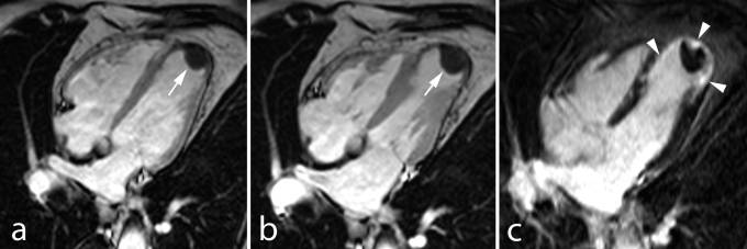
Cardiac magnetic resonance imaging (MRI) has emerged as a prime player in the clinical and preclinical detection of ischemic heart disease (IHD) as well in the prognosis assessment by offering a comprehensive approach for all spectrums of coronary artery disease (CAD) patients. The aim of this review is to provide the reader a state-of-the art on how the newest cardiac MRI techniques can be used to study IHD patients. In patients with suspected/stable CAD, functional and perfusion imaging both at rest and during vasodilatatory stress (adenosine, dypiridamole)/dobutamine stress can accurately depict ischemic myocardium secondary to significant coronary artery stenosis. In patients with acute MI, MRI is a robust tool for differentiating and sizing the jeopardized and the infarcted myocardium by using a combination of functional, edema, perfusion and Gd contrast imaging. Moreover, important prognostic factors like myocardial salvage, the presence of microvascular obstruction (MVO), post reperfusion myocardial hemorrhage, RV involvement and infarct related complications can be assessed in the same examination. In patients with chronic ischemic cardiomyopathy, the role of the MRI extends from diagnosis by means of Gadolinium contrast scar imaging to therapy and prognosis by functional assessment and viability testing with rest and dobutamine stress imaging. In all the circumstances mentioned, MRI derived information has been proven valuable in every day clinical decision making and prognosis assessment. Thus, MRI is becoming more and more an accepted alternative to other imaging modalities both in the acute and chronic setting.
心脏磁共振成像(MRI)已成为临床和临床前检测缺血性心脏病(IHD)以及通过为所有类型的冠状动脉疾病(CAD)患者提供全面方法进行预后评估的主要手段。本综述的目的是向读者介绍最新的心脏MRI技术如何用于研究IHD患者。在疑似/稳定CAD患者中,静息和血管扩张应激(腺苷、双嘧达莫)/多巴酚丁胺应激下的功能和灌注成像都可以准确描绘继发于严重冠状动脉狭窄的缺血心肌。在急性心肌梗死患者中,MRI是一种强大的工具,通过结合功能、水肿、灌注和钆对比成像来区分和测量濒危心肌和梗死心肌的大小。此外,在同一项检查中可以评估重要的预后因素,如心肌挽救、微血管阻塞(MVO)的存在、再灌注后心肌出血、右心室受累和梗死相关并发症。在慢性缺血性心肌病患者中,MRI的作用从通过钆对比瘢痕成像进行诊断扩展到通过静息和多巴酚丁胺应激成像进行功能评估和存活测试来进行治疗和预后评估。在上述所有情况下,MRI获得的信息已被证明在日常临床决策和预后评估中具有价值。因此,在急性和慢性情况下,MRI越来越成为其他成像方式的可接受替代方法。