Department of Ophthalmology, University of Ulm, Prittwitzstrasse 43, D-89075 Ulm, Germany.
Br J Ophthalmol. 2012 Jul;96(7):1023-8. doi: 10.1136/bjophthalmol-2012-301677. Epub 2012 Apr 26.
Retinal endothelial cells are crucially involved in the genesis of diabetic retinopathy which is treated with vascular endothelial growth factor (VEGF) inhibitors. Of these, ranibizumab can completely restore VEGF-induced effects on immortalised bovine retinal endothelial cells (iBREC). In most experiments supporting diabetic retinopathy therapy with bevacizumab, only non-retinal EC or retinal pigment epithelial cells have been used. Also, bevacizumab but not ranibizumab can accumulate in retinal pigment epithelial cells.
To investigate the effects of bevacizumab on VEGF-induced changes of iBREC properties and potential uptake and accumulation of both inhibitors.
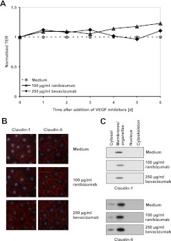
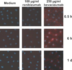
Uptake of VEGF inhibitors by iBREC with or without pretreatment with VEGF(165) was visualised by immunofluorescence staining and western blot analyses. Measured transendothelial resistance (TER) of iBREC (±VEGF(165)) showed effects on permeability, indicated also by the western blot-determined tight junction protein claudin-1. The influence of bevacizumab on proliferation and migration of iBREC was studied in the presence and absence of VEGF(165).
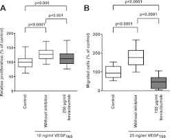
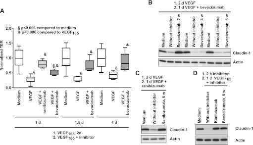
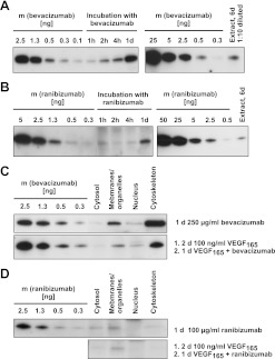
Bevacizumab strongly inhibited VEGF-stimulated and basal migration, but was less efficient than ranibizumab in inhibiting VEGF-induced proliferation or restoring the VEGF-induced decrease of TER and claudin-1. This ability was completely lost after storage of bevacizumab for 4 weeks at 4°C. Ranibizumab and bevacizumab were detectable in whole cell extracts after treatment for at least 1 h; bevacizumab accumulated during prolonged treatment. Ranibizumab was found in the membrane/organelle fraction, whereas bevacizumab was associated with the cytoskeleton.
Both inhibitors had similar effects on retinal endothelial cells; however, some differences were recognised. Although barrier properties were not affected by internalised bevacizumab in vitro, potential adverse effects due to accumulation after repetitive intravitreal injections remain to be investigated.
视网膜内皮细胞在糖尿病性视网膜病变的发生中起着至关重要的作用,该疾病采用血管内皮生长因子 (VEGF) 抑制剂进行治疗。其中,雷珠单抗可完全恢复 VEGF 对永生化牛视网膜内皮细胞 (iBREC) 的诱导作用。在支持贝伐单抗治疗糖尿病性视网膜病变的大多数实验中,仅使用了非视网膜 EC 或视网膜色素上皮细胞。此外,贝伐单抗而非雷珠单抗可在视网膜色素上皮细胞中积累。
研究贝伐单抗对 iBREC 中 VEGF 诱导的变化以及两种抑制剂的潜在摄取和积累的影响。
通过免疫荧光染色和 Western blot 分析观察 iBREC 对 VEGF 抑制剂的摄取,或有无 VEGF(165)预处理。iBREC(±VEGF(165)) 的跨内皮电阻 (TER) 测量表明对通透性有影响,Western blot 测定的紧密连接蛋白 Claudin-1 也表明了这一点。研究了在存在和不存在 VEGF(165)的情况下,贝伐单抗对 iBREC 增殖和迁移的影响。
贝伐单抗强烈抑制 VEGF 刺激和基础迁移,但在抑制 VEGF 诱导的增殖或恢复 VEGF 诱导的 TER 和 Claudin-1 下降方面的效率低于雷珠单抗。这种能力在 4°C 下储存贝伐单抗 4 周后完全丧失。雷珠单抗和贝伐单抗在治疗至少 1 小时后可在全细胞提取物中检测到;贝伐单抗在长时间治疗过程中积累。雷珠单抗存在于膜/细胞器部分,而贝伐单抗与细胞骨架相关。
两种抑制剂对视网膜内皮细胞均有相似的作用;然而,也发现了一些差异。尽管在体外,内化的贝伐单抗对屏障特性没有影响,但仍需研究重复玻璃体内注射后因积累而产生的潜在不良影响。