• 文献检索
  • 文档翻译
  • 深度研究
  • 学术资讯
  • Suppr Zotero 插件Zotero 插件
  • 邀请有礼
  • 套餐&价格
  • 历史记录
应用&插件
Suppr Zotero 插件Zotero 插件浏览器插件Mac 客户端Windows 客户端微信小程序
定价
高级版会员购买积分包购买API积分包
服务
文献检索文档翻译深度研究API 文档MCP 服务
关于我们
关于 Suppr公司介绍联系我们用户协议隐私条款
关注我们

Suppr 超能文献

核心技术专利:CN118964589B侵权必究
粤ICP备2023148730 号-1Suppr @ 2026

文献检索

告别复杂PubMed语法,用中文像聊天一样搜索,搜遍4000万医学文献。AI智能推荐,让科研检索更轻松。

立即免费搜索

文件翻译

保留排版,准确专业,支持PDF/Word/PPT等文件格式,支持 12+语言互译。

免费翻译文档

深度研究

AI帮你快速写综述,25分钟生成高质量综述,智能提取关键信息,辅助科研写作。

立即免费体验

大鼠实验性蛛网膜下腔出血后最初6小时内,脑水肿形成与灌注不足相关。

Brain edema formation correlates with perfusion deficit during the first six hours after experimental subarachnoid hemorrhage in rats.

作者信息

Westermaier Thomas, Stetter Christian, Raslan Furat, Vince Giles Hamilton, Ernestus Ralf-Ingo

机构信息

Department of Neurosurgery, University of Würzburg, Josef-Schneider-Str, 11, Würzburg, 97080, Germany.

出版信息

Exp Transl Stroke Med. 2012 Jul 13;4(1):8. doi: 10.1186/2040-7378-4-8.

DOI:10.1186/2040-7378-4-8
PMID:22551223
原文链接:https://pmc.ncbi.nlm.nih.gov/articles/PMC3398845/
Abstract

BACKGROUND

Severe brain edema is observed in a number of patients suffering from subarachnoid hemorrhage (SAH). Little is known about its pathogenesis and time-course in the first hours after SAH. This study was performed to investigate the development of brain edema and its correlation with brain perfusion after experimental SAH.

METHODS

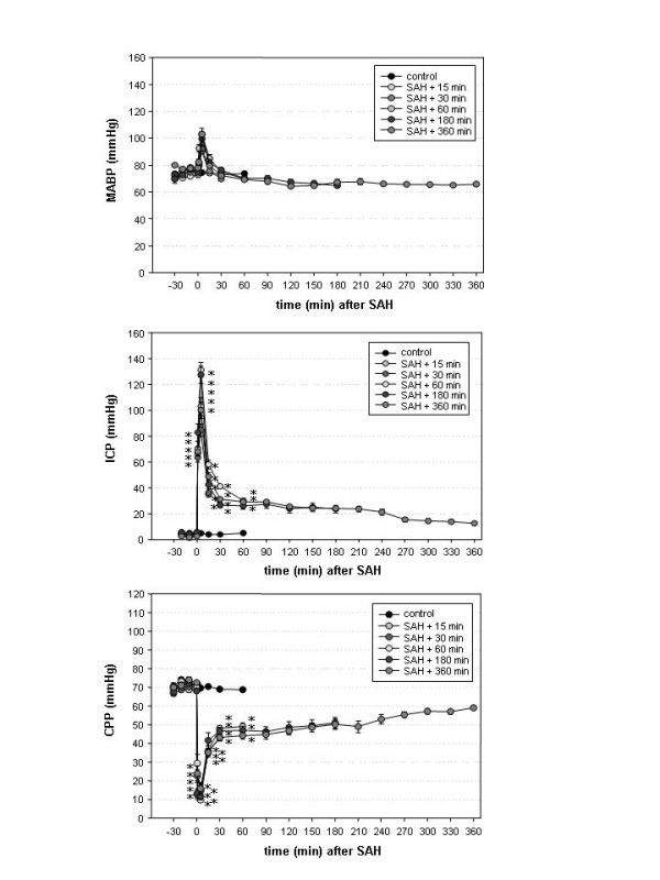
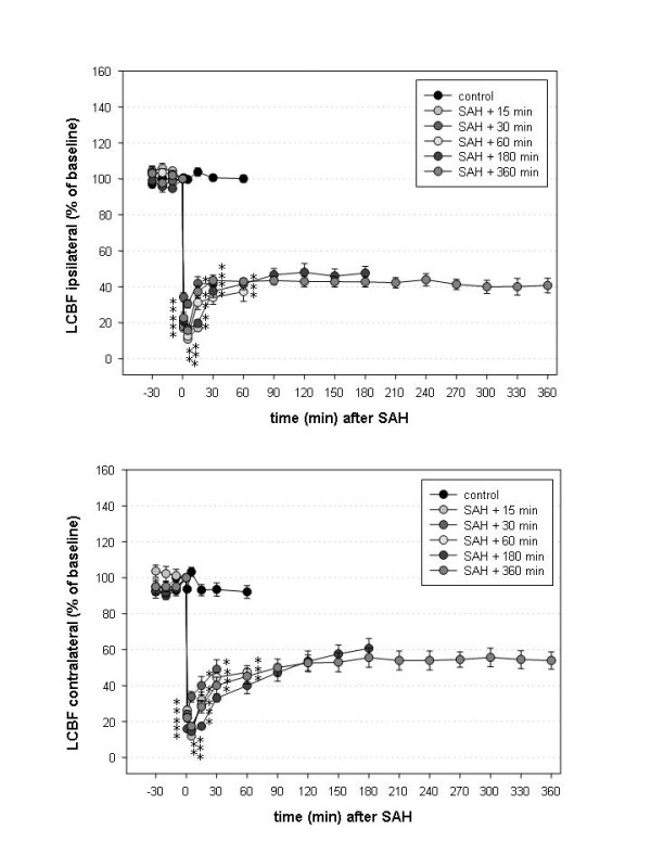
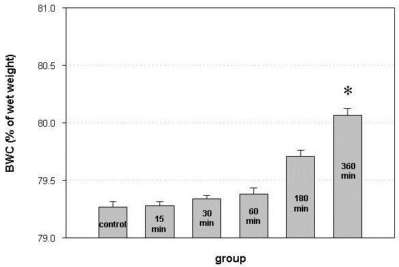
Male Sprague-Dawley rats, randomly assigned to one of six groups (n = 8), were subjected to SAH using the endovascular filament model or underwent a sham operation. Animals were sacrificed 15, 30, 60, 180 or 360 minutes after SAH. Intracranial pressure (ICP), mean arterial blood pressure (MABP), cerebral perfusion pressure (CPP) and bilateral local cerebral blood flow (LCBF) were continuously measured. Brain water content (BWC) was determined by the wet/dry-weight method.

RESULTS

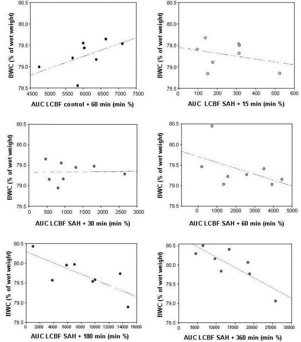
After SAH, CPP and LCBF rapidly decreased. The decline of LCBF markedly exceeded the decline of CPP and persisted until the end of the observation period. BWC continuously increased. A significant correlation was observed between the BWC and the extent of the perfusion deficit in animals sacrificed after 180 and 360 minutes.

CONCLUSIONS

The significant correlation with the perfusion deficit after SAH suggests that the development of brain edema is related to the extent of ischemia and acute vasoconstriction in the first hours after SAH.

摘要

背景

在许多蛛网膜下腔出血(SAH)患者中观察到严重脑水肿。关于其在SAH后最初几小时内的发病机制和时间进程知之甚少。本研究旨在探讨实验性SAH后脑水肿的发展及其与脑灌注的相关性。

方法

将雄性Sprague-Dawley大鼠随机分为六组(每组n = 8),采用血管内丝线模型进行SAH或进行假手术。在SAH后15、30、60、180或360分钟处死动物。连续测量颅内压(ICP)、平均动脉血压(MABP)、脑灌注压(CPP)和双侧局部脑血流量(LCBF)。采用湿/干重法测定脑含水量(BWC)。

结果

SAH后,CPP和LCBF迅速下降。LCBF的下降明显超过CPP的下降,并持续到观察期结束。BWC持续增加。在180和360分钟后处死的动物中,观察到BWC与灌注缺损程度之间存在显著相关性。

结论

SAH后与灌注缺损的显著相关性表明,脑水肿的发展与SAH后最初几小时内的缺血程度和急性血管收缩有关。

https://cdn.ncbi.nlm.nih.gov/pmc/blobs/26d2/3398845/95c75ac24a36/2040-7378-4-8-4.jpg
https://cdn.ncbi.nlm.nih.gov/pmc/blobs/26d2/3398845/8859f590781e/2040-7378-4-8-1.jpg
https://cdn.ncbi.nlm.nih.gov/pmc/blobs/26d2/3398845/0734e8046e3a/2040-7378-4-8-2.jpg
https://cdn.ncbi.nlm.nih.gov/pmc/blobs/26d2/3398845/cdc4f6c6331e/2040-7378-4-8-3.jpg
https://cdn.ncbi.nlm.nih.gov/pmc/blobs/26d2/3398845/95c75ac24a36/2040-7378-4-8-4.jpg

相似文献

1
Brain edema formation correlates with perfusion deficit during the first six hours after experimental subarachnoid hemorrhage in rats.大鼠实验性蛛网膜下腔出血后最初6小时内,脑水肿形成与灌注不足相关。
Exp Transl Stroke Med. 2012 Jul 13;4(1):8. doi: 10.1186/2040-7378-4-8.
2
Acute vasoconstriction: decrease and recovery of cerebral blood flow after various intensities of experimental subarachnoid hemorrhage in rats.急性血管收缩:大鼠不同强度实验性蛛网膜下腔出血后脑血流量的减少与恢复
J Neurosurg. 2009 May;110(5):996-1002. doi: 10.3171/2008.8.JNS08591.
3
Acute and delayed vasoconstriction after subarachnoid hemorrhage: local cerebral blood flow, histopathology, and morphology in the rat basilar artery.蛛网膜下腔出血后的急性和延迟性血管收缩:大鼠基底动脉的局部脑血流量、组织病理学和形态学
Arch Physiol Biochem. 2001 Apr;109(2):145-53. doi: 10.1076/apab.109.2.145.4267.
4
Clazosentan, an endothelin receptor antagonist, prevents early hypoperfusion during the acute phase of massive experimental subarachnoid hemorrhage: a laser Doppler flowmetry study in rats.内皮素受体拮抗剂克拉生坦可预防大鼠大规模实验性蛛网膜下腔出血急性期的早期低灌注:一项激光多普勒血流仪研究
J Neurosurg. 2008 Dec;109(6):1134-40. doi: 10.3171/JNS.2008.109.12.1134.
5
Acute vasoconstriction after subarachnoid hemorrhage.蛛网膜下腔出血后的急性血管收缩
Neurosurgery. 1998 Feb;42(2):352-60; discussion 360-2. doi: 10.1097/00006123-199802000-00091.
6
Time-course of cerebral perfusion and tissue oxygenation in the first 6 h after experimental subarachnoid hemorrhage in rats.大鼠实验性蛛网膜下腔出血后最初6小时内脑灌注和组织氧合的时间进程。
J Cereb Blood Flow Metab. 2009 Apr;29(4):771-9. doi: 10.1038/jcbfm.2008.169. Epub 2009 Jan 21.
7
Early Antiinflammatory Therapy Attenuates Brain Damage After Sah in Rats.早期抗炎治疗可减轻大鼠蛛网膜下腔出血后的脑损伤。
Transl Neurosci. 2019 Apr 23;10:104-111. doi: 10.1515/tnsci-2019-0018. eCollection 2019.
8
Cortical blood flow and cerebral perfusion pressure in a new noncraniotomy model of subarachnoid hemorrhage in the rat.大鼠蛛网膜下腔出血新的非开颅模型中的皮质血流和脑灌注压
Stroke. 1995 Jun;26(6):1086-91; discussion 1091-2. doi: 10.1161/01.str.26.6.1086.
9
First Description of Reduced Pyruvate Dehydrogenase Enzyme Activity Following Subarachnoid Hemorrhage (SAH).蛛网膜下腔出血(SAH)后丙酮酸脱氢酶活性降低的首次描述。
Front Neurosci. 2017 Feb 14;11:37. doi: 10.3389/fnins.2017.00037. eCollection 2017.
10
Acute reaction of arterial blood vessels after experimental subarachnoid hemorrhage - An in vivo microscopic study.实验性蛛网膜下腔出血后动脉血管的急性反应——活体显微镜研究。
J Neurol Sci. 2019 Jan 15;396:172-177. doi: 10.1016/j.jns.2018.11.007. Epub 2018 Nov 8.

引用本文的文献

1
Matrine exerts its neuroprotective effects by modulating multiple neuronal pathways.苦参碱通过调节多种神经元通路发挥其神经保护作用。
Metab Brain Dis. 2023 Jun;38(5):1471-1499. doi: 10.1007/s11011-023-01214-6. Epub 2023 Apr 27.
2
Lesional and perilesional tissue characterization by automated image processing in a novel gyrencephalic animal model of peracute intracerebral hemorrhage.在一种新的类脑回动物急性脑出血模型中,通过自动化图像处理进行病灶和病灶周围组织特征描述。
J Cereb Blood Flow Metab. 2019 Dec;39(12):2521-2535. doi: 10.1177/0271678X18802119. Epub 2018 Sep 21.
3
Mechanisms of Global Cerebral Edema Formation in Aneurysmal Subarachnoid Hemorrhage.

本文引用的文献

1
Early micro vascular changes after subarachnoid hemorrhage.蛛网膜下腔出血后的早期微血管变化。
Acta Neurochir Suppl. 2011;110(Pt 1):49-55. doi: 10.1007/978-3-7091-0353-1_9.
2
The temporal profile of cerebral blood flow and tissue metabolites indicates sustained metabolic depression after experimental subarachnoid hemorrhage in rats.脑血流和组织代谢物的时程变化表明,在大鼠实验性蛛网膜下腔出血后存在持续的代谢抑制。
Neurosurgery. 2011 Jan;68(1):223-9; discussion 229-30. doi: 10.1227/NEU.0b013e3181fe23c1.
3
The neurovascular unit, matrix proteases, and innate inflammation.
动脉瘤性蛛网膜下腔出血中全脑水肿形成的机制
Neurocrit Care. 2017 Apr;26(2):301-310. doi: 10.1007/s12028-016-0354-7.
4
Propofol Attenuates Early Brain Injury After Subarachnoid Hemorrhage in Rats.丙泊酚减轻大鼠蛛网膜下腔出血后的早期脑损伤。
J Mol Neurosci. 2015 Dec;57(4):538-45. doi: 10.1007/s12031-015-0634-2. Epub 2015 Sep 5.
5
Arginine-Vasopressin Receptor Blocker Conivaptan Reduces Brain Edema and Blood-Brain Barrier Disruption after Experimental Stroke in Mice.精氨酸加压素受体阻滞剂考尼伐坦可减轻小鼠实验性中风后的脑水肿并减少血脑屏障破坏。
PLoS One. 2015 Aug 14;10(8):e0136121. doi: 10.1371/journal.pone.0136121. eCollection 2015.
6
Early brain injury linearly correlates with reduction in cerebral perfusion pressure during the hyperacute phase of subarachnoid hemorrhage.在蛛网膜下腔出血超急性期,早期脑损伤与脑灌注压降低呈线性相关。
Intensive Care Med Exp. 2014 Dec;2(1):30. doi: 10.1186/s40635-014-0030-1. Epub 2014 Nov 30.
7
Controversies and evolving new mechanisms in subarachnoid hemorrhage.蛛网膜下腔出血的争议与新机制的演变
Prog Neurobiol. 2014 Apr;115:64-91. doi: 10.1016/j.pneurobio.2013.09.002. Epub 2013 Sep 25.
8
Pathophysiological Role of Global Cerebral Ischemia following Subarachnoid Hemorrhage: The Current Experimental Evidence.蛛网膜下腔出血后全脑缺血的病理生理作用:当前的实验证据
Stroke Res Treat. 2013;2013:651958. doi: 10.1155/2013/651958. Epub 2013 Jun 12.
神经血管单元、基质蛋白酶和固有炎症。
Ann N Y Acad Sci. 2010 Oct;1207:46-9. doi: 10.1111/j.1749-6632.2010.05760.x.
4
Early global brain oedema in relation to clinical admission parameters and outcome in patients with aneurysmal subarachnoid haemorrhage.与动脉瘤性蛛网膜下腔出血患者入院参数和预后相关的早期全脑水肿。
Acta Neurochir (Wien). 2010 Sep;152(9):1527-33; discussion 1533. doi: 10.1007/s00701-010-0684-8. Epub 2010 May 22.
5
Inflammatory mechanisms in ischemic stroke: role of inflammatory cells.缺血性脑卒中的炎症机制:炎症细胞的作用。
J Leukoc Biol. 2010 May;87(5):779-89. doi: 10.1189/jlb.1109766. Epub 2010 Feb 3.
6
Brain edema formation and neurological impairment after subarachnoid hemorrhage in rats. Laboratory investigation.大鼠蛛网膜下腔出血后脑水肿形成及神经功能损害。实验室研究。
J Neurosurg. 2009 Nov;111(5):988-94. doi: 10.3171/2009.3.JNS08412.
7
Time-course of cerebral perfusion and tissue oxygenation in the first 6 h after experimental subarachnoid hemorrhage in rats.大鼠实验性蛛网膜下腔出血后最初6小时内脑灌注和组织氧合的时间进程。
J Cereb Blood Flow Metab. 2009 Apr;29(4):771-9. doi: 10.1038/jcbfm.2008.169. Epub 2009 Jan 21.
8
Acute vasoconstriction: decrease and recovery of cerebral blood flow after various intensities of experimental subarachnoid hemorrhage in rats.急性血管收缩:大鼠不同强度实验性蛛网膜下腔出血后脑血流量的减少与恢复
J Neurosurg. 2009 May;110(5):996-1002. doi: 10.3171/2008.8.JNS08591.
9
Clazosentan, an endothelin receptor antagonist, prevents early hypoperfusion during the acute phase of massive experimental subarachnoid hemorrhage: a laser Doppler flowmetry study in rats.内皮素受体拮抗剂克拉生坦可预防大鼠大规模实验性蛛网膜下腔出血急性期的早期低灌注:一项激光多普勒血流仪研究
J Neurosurg. 2008 Dec;109(6):1134-40. doi: 10.3171/JNS.2008.109.12.1134.
10
Potential mechanisms and clinical significance of global cerebral edema following aneurysmal subarachnoid hemorrhage.动脉瘤性蛛网膜下腔出血后全脑水肿的潜在机制及临床意义
Neurosurg Focus. 2007 May 15;22(5):E7. doi: 10.3171/foc.2007.22.5.8.