Department of Pathology, Microbiology and Immunology, Vanderbilt University School of Medicine, Nashville, TN 37232-2363, USA.
J Clin Immunol. 2012 Oct;32(5):1129-40. doi: 10.1007/s10875-012-9700-5. Epub 2012 May 3.
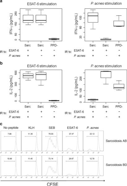
Sarcoidosis is a non-caseating granulomatous disease for which a role for infectious antigens continues to strengthen. Recent studies have reported molecular evidence of mycobacteria or propionibacteria. We assessed for immune responses against mycobacterial and propionibacterial antigens in sarcoidosis bronchoalveolar lavage (BAL) using flow cytometry, and localized signals consistent with microbial antigens with sarcoidosis specimens, using matrix-assisted laser desorption ionization imaging mass spectrometry (MALDI-IMS).
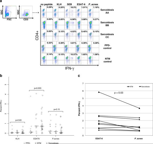
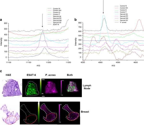
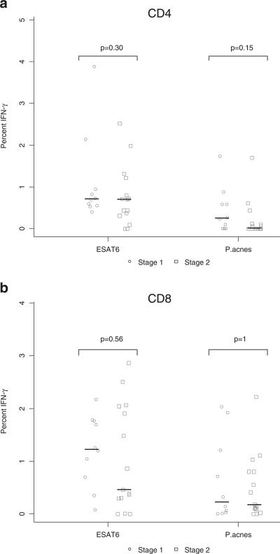
BAL cells from 27 sarcoidosis, 14 PPD- controls, and 9 subjects with nontuberculosis mycobacterial (NTM) infections were analyzed for production of IFN-γ after stimulation with mycobacterial ESAT-6 and Propionibacterium acnes proteins. To complement the immunological data, MALDI-IMS was performed to localize ESAT-6 and Propionibacterium acnes signals within sarcoidosis and control specimens.
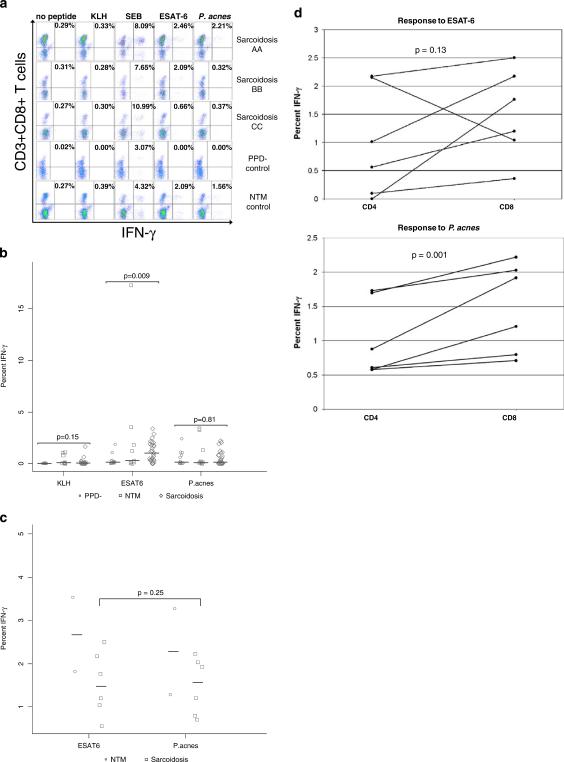
CD4+ immunologic analysis for mycobacteria was positive in 17/27 sarcoidosis subjects, compared to 2/14 PPD- subjects, and 5/9 NTM subjects (p = 0.008 and p = 0.71 respectively, Fisher's exact test). There was no significant difference for recognition of P. acnes, which occurred only in sarcoidosis subjects that also recognized ESAT-6. Similar results were also observed for the CD8+ immunologic analysis. MALDI-IMS localized signals consistent with ESAT-6 only within sites of granulomatous inflammation, whereas P. acnes signals were distributed throughout the specimen.
MALDI-IMS localizes signals consistent with ESAT-6 to sarcoidosis granulomas, whereas no specific localization of P. acnes signals is detected. Immune responses against both mycobacterial and P. acnes are present within sarcoidosis BAL, but only mycobacterial signals are distinct from disease controls. These immunologic and molecular investigations support further investigation of the microbial community within sarcoidosis granulomas.
结节病是一种非干酪样肉芽肿性疾病,其感染抗原的作用持续增强。最近的研究报告了分枝杆菌或丙酸杆菌的分子证据。我们使用流式细胞术评估结节病支气管肺泡灌洗液(BAL)中针对分枝杆菌和丙酸杆菌抗原的免疫反应,并使用基质辅助激光解吸电离成像质谱(MALDI-IMS)在结节病标本中定位与微生物抗原一致的信号。
分析 27 例结节病、14 例 PPD-对照和 9 例非结核分枝杆菌(NTM)感染患者的 BAL 细胞,在刺激后产生 IFN-γ分枝杆菌 ESAT-6 和丙酸杆菌蛋白。为了补充免疫学数据,进行 MALDI-IMS 以在结节病和对照标本中定位 ESAT-6 和丙酸杆菌信号。
与 14 例 PPD-对照和 9 例 NTM 对照相比,17/27 例结节病患者的分枝杆菌 CD4+免疫分析为阳性(p=0.008 和 p=0.71,Fisher 精确检验)。对 P. acnes 的识别没有显着差异,仅在识别 ESAT-6 的结节病患者中发生。CD8+免疫分析也观察到类似的结果。MALDI-IMS 仅在肉芽肿炎症部位定位与 ESAT-6 一致的信号,而丙酸杆菌信号分布在整个标本中。
MALDI-IMS 将与 ESAT-6 一致的信号定位到结节病肉芽肿中,而未检测到丙酸杆菌信号的特定定位。在结节病 BAL 中存在针对分枝杆菌和丙酸杆菌的免疫反应,但只有分枝杆菌信号与疾病对照不同。这些免疫学和分子研究支持进一步研究结节病肉芽肿中的微生物群落。