Division of Cell Biology, Kihara Institute for Biological Research, Yokohama City University, Yokohama, Japan.
PLoS One. 2012;7(5):e35546. doi: 10.1371/journal.pone.0035546. Epub 2012 May 1.
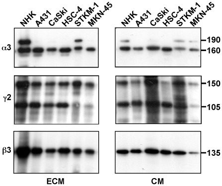
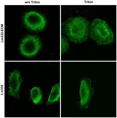
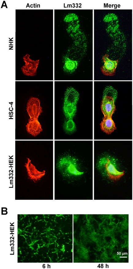
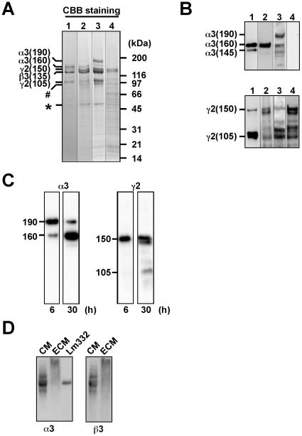
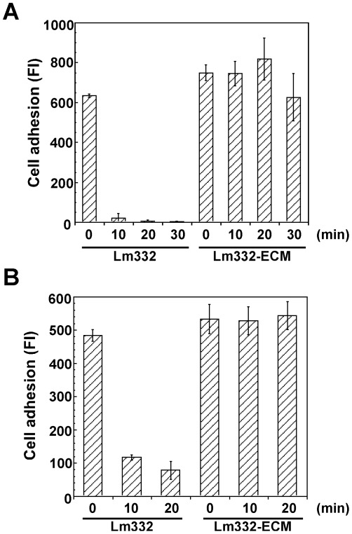
Laminin-332 (α3ß3γ2) (Lm332) supports the stable anchoring of basal keratinocytes to the epidermal basement membrane, while it functions as a motility factor for wound healing and cancer invasion. To understand these contrasting activities of Lm332, we investigated Lm332 matrices deposited by normal human keratinocytes and other Lm332-expressing cell lines. All types of the cells efficiently deposited Lm332 on the culture plates in specific patterns. On the contrary, laminins containing laminin ß1 and/or γ1 chains, such as Lm511 and Lm311, were not deposited on the culture plates even if secreted into culture medium. The Lm332 deposition was not inhibited by function-blocking antibodies to the α3 and α6 integrins but was inhibited by sodium selenate, suggesting that sulfated glycosaminoglycans on cell surface, e.g. heparan sulfate proteoglycans, might be involved in the process. HEK293 cells overexpressing exogenous Lm332 (Lm332-HEK) almost exclusively deposited Lm332 on the plates. The deposited Lm332 matrix showed a mesh-like network structure as analyzed by electron microscopy, suggesting that Lm332 was highly polymerized. When biological activity was analyzed, the Lm332 matrix rather suppressed the migration of keratinocytes as compared with purified Lm332, which highly promoted the cell migration. The Lm332 matrix supported adhesion of keratinocytes much more strongly and stably than purified Lm332. Integrin α3ß1 bound to the Lm332 matrix at a three times higher level than purified Lm332. Normal keratinocytes prominently showed integrin α6ß4-containing, hemidesmosome-like structures on the Lm332 matrix but not on the purified one. These results indicate that the polymerized Lm332 matrix supports stable cell adhesion by interacting with both integrin α6ß4 and α3ß1, whereas unassembled soluble Lm332 supports cell migration.
层粘连蛋白-332(α3ß3γ2)(Lm332)支持基底角质形成细胞与表皮基底膜的稳定锚定,同时作为伤口愈合和癌症侵袭的运动因子发挥作用。为了了解 Lm332 的这些对比活性,我们研究了正常人类角质形成细胞和其他表达 Lm332 的细胞系沉积的 Lm332 基质。所有类型的细胞都能有效地在培养板上以特定的模式沉积 Lm332。相反,含有层粘连蛋白β1 和/或γ1 链的层粘连蛋白,如 Lm511 和 Lm311,即使分泌到培养基中也不会沉积在培养板上。Lm332 的沉积不受针对α3 和α6 整联蛋白的功能阻断抗体的抑制,但被亚硒酸钠抑制,表明细胞表面上的硫酸化糖胺聚糖,例如硫酸乙酰肝素蛋白聚糖,可能参与该过程。过表达外源 Lm332 的 HEK293 细胞(Lm332-HEK)几乎只在平板上沉积 Lm332。电子显微镜分析显示,沉积的 Lm332 基质呈现网状网络结构,表明 Lm332 高度聚合。当分析生物活性时,与高度促进细胞迁移的纯化 Lm332 相比,Lm332 基质对角质形成细胞的迁移抑制作用更强。Lm332 基质对角质形成细胞的黏附支持作用比纯化的 Lm332 更强且更稳定。整合素α3ß1 与 Lm332 基质的结合水平比纯化的 Lm332 高三倍。正常角质形成细胞在 Lm332 基质上明显显示出含有整合素α6ß4 的半桥粒样结构,但在纯化的 Lm332 上则没有。这些结果表明,聚合的 Lm332 基质通过与整合素α6ß4 和α3ß1 相互作用来支持稳定的细胞黏附,而未组装的可溶性 Lm332 则支持细胞迁移。