Department of Physiological Science, Universidade Federal de Goiás, Goiânia, Goiás, Brazil.
PLoS One. 2012;7(5):e37587. doi: 10.1371/journal.pone.0037587. Epub 2012 May 21.
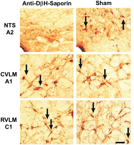
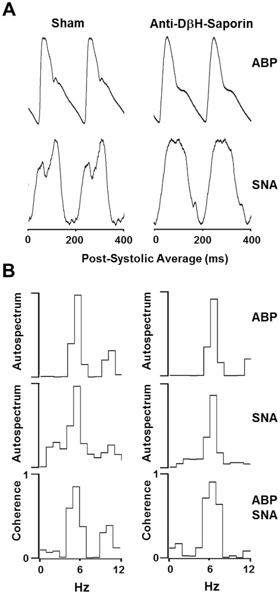
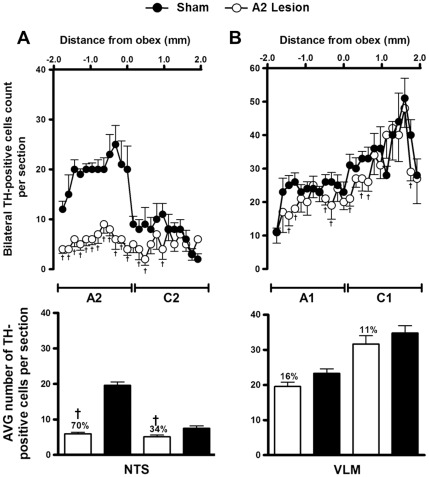
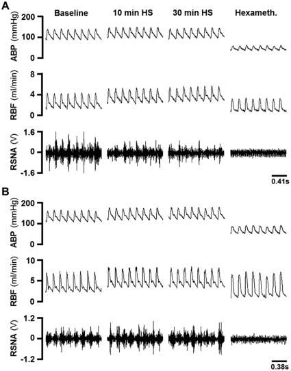
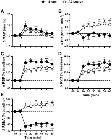
Renal vasodilation and sympathoinhibition are recognized responses induced by hypernatremia, but the central neural pathways underlying such responses are not yet entirely understood. Several findings suggest that A2 noradrenergic neurons, which are found in the nucleus of the solitary tract (NTS), play a role in the pathways that contribute to body fluid homeostasis and cardiovascular regulation. The purpose of this study was to determine the effects of selective lesions of A2 neurons on the renal vasodilation and sympathoinhibition induced by hypertonic saline (HS) infusion. Male Wistar rats (280-350 g) received an injection into the NTS of anti-dopamine-beta-hydroxylase-saporin (A2 lesion; 6.3 ng in 60 nl; n = 6) or free saporin (sham; 1.3 ng in 60 nl; n = 7). Two weeks later, the rats were anesthetized (urethane 1.2 g⋅kg(-1) b.wt., i.v.) and the blood pressure, renal blood flow (RBF), renal vascular conductance (RVC) and renal sympathetic nerve activity (RSNA) were recorded. In sham rats, the HS infusion (3 M NaCl, 1.8 ml⋅kg(-1) b.wt., i.v.) induced transient hypertension (peak at 10 min after HS; 9±2.7 mmHg) and increases in the RBF and RVC (141±7.9% and 140±7.9% of baseline at 60 min after HS, respectively). HS infusion also decreased the RSNA (-45±5.0% at 10 min after HS) throughout the experimental period. In the A2-lesioned rats, the HS infusion induced transient hypertension (6±1.4 mmHg at 10 min after HS), as well as increased RBF and RVC (133±5.2% and 134±6.9% of baseline at 60 min after HS, respectively). However, in these rats, the HS failed to reduce the RSNA (115±3.1% at 10 min after HS). The extent of the catecholaminergic lesions was confirmed by immunocytochemistry. These results suggest that A2 noradrenergic neurons are components of the neural pathways regulating the composition of the extracellular fluid compartment and are selectively involved in hypernatremia-induced sympathoinhibition.
肾脏血管舒张和交感神经抑制是高钠血症引起的公认反应,但这种反应的中枢神经通路尚未完全了解。有几项研究结果表明,位于孤束核(NTS)中的 A2 去甲肾上腺素能神经元在参与体液平衡和心血管调节的通路中发挥作用。本研究的目的是确定选择性 A2 神经元损伤对高渗盐水(HS)输注引起的肾脏血管舒张和交感神经抑制的影响。雄性 Wistar 大鼠(280-350g)接受 NTS 内注射抗多巴胺-β-羟化酶-相思豆毒素(A2 损伤;6.3ng 在 60nl 中;n=6)或游离相思豆毒素(假手术;1.3ng 在 60nl 中;n=7)。两周后,大鼠麻醉(乌拉坦 1.2g/kg 体重,静脉内),记录血压、肾血流量(RBF)、肾血管传导率(RVC)和肾交感神经活性(RSNA)。在假手术组中,HS 输注(3M NaCl,1.8ml/kg 体重,静脉内)引起短暂性高血压(HS 后 10min 时峰值;9±2.7mmHg)和 RBF 和 RVC 增加(HS 后 60min 时分别为基础值的 141±7.9%和 140±7.9%)。HS 输注还在整个实验期间降低了 RSNA(HS 后 10min 时为-45±5.0%)。在 A2 损伤大鼠中,HS 输注引起短暂性高血压(HS 后 10min 时为 6±1.4mmHg),以及 RBF 和 RVC 增加(HS 后 60min 时分别为基础值的 133±5.2%和 134±6.9%)。然而,在这些大鼠中,HS 未能降低 RSNA(HS 后 10min 时为 115±3.1%)。通过免疫细胞化学证实了儿茶酚胺能神经元损伤的程度。这些结果表明,A2 去甲肾上腺素能神经元是调节细胞外液成分的神经通路的组成部分,并且选择性地参与高钠血症引起的交感神经抑制。