Institute of Cell Biology and Neurobiology, Center for Anatomy, Charité - Universitätsmedizin Berlin, Charitéplatz 1, 10117, Berlin, Germany.
Neural Dev. 2012 Jun 13;7:21. doi: 10.1186/1749-8104-7-21.
During neocortical development, multiple voltage- and ligand-gated ion channels are differentially expressed in neurons thereby shaping their intrinsic electrical properties. One of these voltage-gated ion channels, the hyperpolarization-activated cyclic nucleotide-gated (HCN) channel and its current I(h), is an important regulator of neuronal excitability. Thus far, studies on an early I(h) appearance in rodent neocortex are missing or conflicting. Therefore, we focused our study on perinatal neocortical I(h) and its properties.
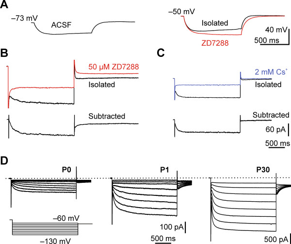
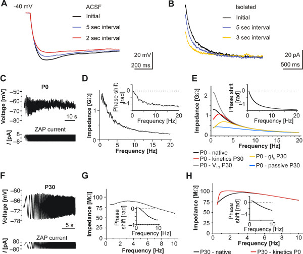
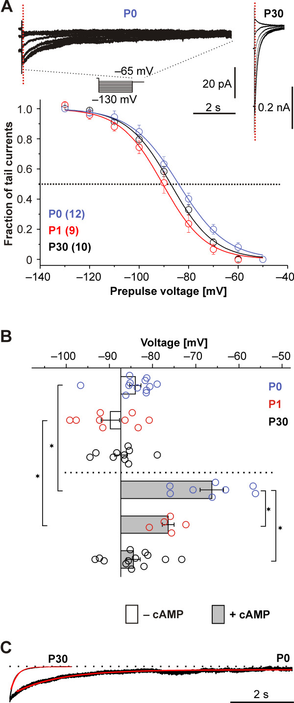
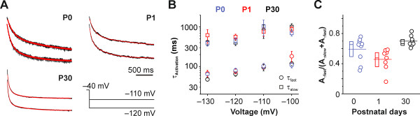
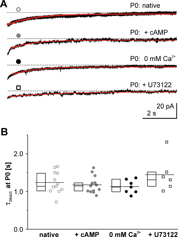
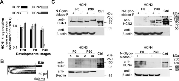
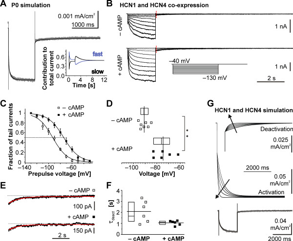
In the perinatal rat neocortex we observed a rapid increase in the number of neurons exhibiting I(h). Perinatal I(h) had unique properties: first, a pronounced cAMP sensitivity resulting in a marked shift of the voltage sufficient for half-maximum activation of the current towards depolarized voltages and second, an up to 10 times slower deactivation at physiological membrane potentials when compared to the one at postnatal day 30. The combination of these features was sufficient to suppress membrane resonance in our in silico and in vitro experiments. Although all four HCN subunits were present on the mRNA level we only detected HCN4, HCN3 and HCN1 on the protein level at P0. HCN1 protein at P0, however, appeared incompletely processed. At P30 glycosilated HCN1 and HCN2 dominated. By in silico simulations and heterologous co-expression experiments of a 'slow' and a 'fast' I(h) conducting HCN channel subunit in HEK293 cells, we mimicked most characteristics of the native current, pointing to a functional combination of subunit homo- or heteromeres.
Taken together, these data indicate a HCN subunit shift initiated in the first 24 hours after birth and implicate a prominent perinatal role of the phylogenetically older HCN3 and/or HCN4 subunits in the developing neocortex.
在皮质发育过程中,神经元中多种电压门控和配体门控离子通道的表达存在差异,从而形成其固有电特性。这些电压门控离子通道之一,超极化激活环核苷酸门控(HCN)通道及其电流 I(h),是神经元兴奋性的重要调节因子。到目前为止,关于啮齿动物皮质中早期 I(h)出现的研究要么缺失,要么相互矛盾。因此,我们的研究集中在围产期皮质的 I(h)及其特性上。
我们在新生大鼠皮质中观察到具有 I(h)的神经元数量迅速增加。围产期 I(h)具有独特的特性:首先,具有显著的 cAMP 敏感性,导致电流的半激活电压向去极化电压显著偏移;其次,与出生后第 30 天相比,在生理膜电位下,失活速度慢了 10 倍。这些特征的结合足以在我们的体内外实验中抑制膜共振。尽管在 mRNA 水平上存在所有四个 HCN 亚基,但仅在 P0 时在蛋白质水平上检测到 HCN4、HCN3 和 HCN1。然而,P0 时的 HCN1 蛋白似乎未完全加工。在 P30 时,糖基化的 HCN1 和 HCN2 占主导地位。通过在 HEK293 细胞中对“慢”和“快”I(h)传导 HCN 通道亚基进行计算机模拟和异源共表达实验,我们模拟了天然电流的大多数特性,这表明亚基同源或异源二聚体具有功能组合。
综上所述,这些数据表明,在出生后前 24 小时内启动了 HCN 亚基转移,并暗示在发育中的皮质中,进化上较老的 HCN3 和/或 HCN4 亚基具有重要的围产期作用。