School of Biological & Biomedical Sciences, Durham University, Durham DH1 3LE, UK.
Neuroscience. 2012 Sep 6;219(1-2):48-61. doi: 10.1016/j.neuroscience.2012.05.070. Epub 2012 Jun 12.
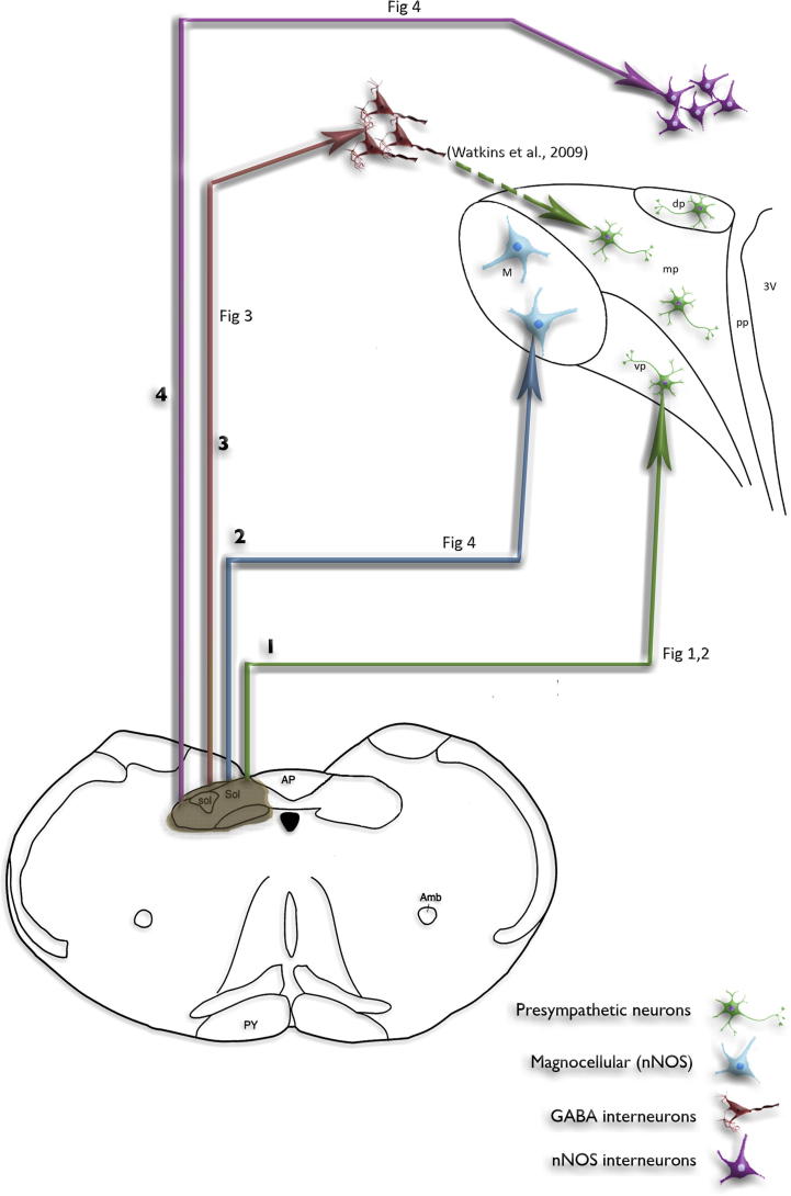
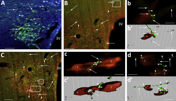
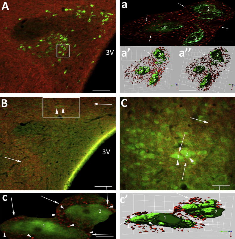
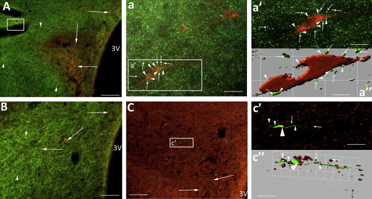
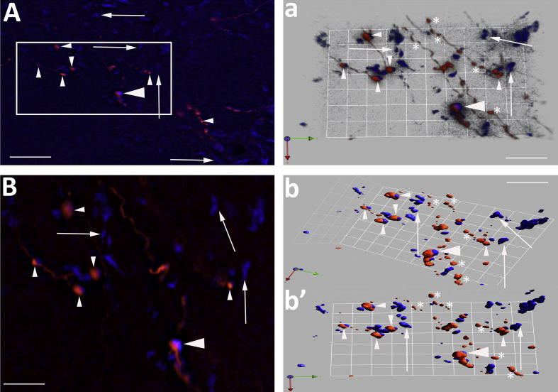
Elevated sympathetic nerve activity, strongly associated with cardiovascular disease, is partly generated from the presympathetic neurons of the paraventricular nucleus of the hypothalamus (PVN). The PVN-presympathetic neurons regulating cardiac and vasomotor sympathetic activity receive information about cardiovascular status from receptors in the heart and circulation. These receptors signal changes via afferent neurons terminating in the nucleus tractus solitarius (NTS), some of which may result in excitation or inhibition of PVN-presympathetic neurons. Understanding the anatomy and neurochemistry of NTS afferent connections within the PVN could provide important clues to the impairment in homeostasis cardiovascular control associated with disease. Transynaptic labelling has shown the presence of neuronal nitric oxide synthase (nNOS)-containing neurons and GABA interneurons that terminate on presympathetic PVN neurons any of which may be the target for NTS afferents. So far NTS connections to these diverse neuronal pools have not been demonstrated and were investigated in this study. Anterograde (biotin dextran amine - BDA) labelling of the ascending projection from the NTS and retrograde (fluorogold - FG or cholera toxin B subunit - CTB) labelling of PVN presympathetic neurons combined with immunohistochemistry for GABA and nNOS was used to identify the terminal neuronal targets of the ascending projection from the NTS. It was shown that NTS afferent terminals are apposed to either PVN-GABA interneurons or to nitric oxide producing neurons or even directly to presympathetic neurons. Furthermore, there was evidence that some NTS axons were positive for vesicular glutamate transporter 2 (vGLUT2). The data provide an anatomical basis for the different functions of cardiovascular receptors that mediate their actions via the NTS-PVN pathways.
交感神经活性升高与心血管疾病密切相关,部分源自下丘脑室旁核 (PVN) 的节前神经元。调节心脏和血管交感神经活动的 PVN-节前神经元从心脏和循环中的受体接收心血管状态信息。这些受体通过终止于孤束核 (NTS) 的传入神经元发出信号变化,其中一些可能导致 PVN-节前神经元兴奋或抑制。了解 NTS 传入连接在 PVN 中的解剖和神经化学,可能为与疾病相关的心血管稳态控制受损提供重要线索。转导标记显示存在神经元型一氧化氮合酶 (nNOS) 包含神经元和 GABA 中间神经元,它们终止于节前 PVN 神经元,其中任何一种都可能成为 NTS 传入的靶标。到目前为止,尚未证明 NTS 与这些不同神经元池的连接,并在本研究中进行了研究。使用 NTS 上行投射的顺行 (生物素葡聚糖胺-BDA) 标记和 PVN 节前神经元的逆行 (荧光金-FG 或霍乱毒素 B 亚单位-CTB) 标记结合 GABA 和 nNOS 的免疫组织化学,用于鉴定 NTS 上行投射的终末神经元靶标。结果表明,NTS 传入末端与 PVN-GABA 中间神经元或产生一氧化氮的神经元或甚至直接与节前神经元相邻。此外,有证据表明,一些 NTS 轴突对囊泡谷氨酸转运体 2 (vGLUT2) 呈阳性。该数据为心血管受体的不同功能提供了解剖学基础,这些受体通过 NTS-PVN 途径介导其作用。