• 文献检索
  • 文档翻译
  • 深度研究
  • 学术资讯
  • Suppr Zotero 插件Zotero 插件
  • 邀请有礼
  • 套餐&价格
  • 历史记录
应用&插件
Suppr Zotero 插件Zotero 插件浏览器插件Mac 客户端Windows 客户端微信小程序
定价
高级版会员购买积分包购买API积分包
服务
文献检索文档翻译深度研究API 文档MCP 服务
关于我们
关于 Suppr公司介绍联系我们用户协议隐私条款
关注我们

Suppr 超能文献

核心技术专利:CN118964589B侵权必究
粤ICP备2023148730 号-1Suppr @ 2026

文献检索

告别复杂PubMed语法,用中文像聊天一样搜索,搜遍4000万医学文献。AI智能推荐,让科研检索更轻松。

立即免费搜索

文件翻译

保留排版,准确专业,支持PDF/Word/PPT等文件格式,支持 12+语言互译。

免费翻译文档

深度研究

AI帮你快速写综述,25分钟生成高质量综述,智能提取关键信息,辅助科研写作。

立即免费体验

正常角膜、未经治疗的扩张性角膜以及先前接受角膜交联治疗的扩张性角膜的上皮厚度之间的相关性;总体上皮厚度是否是早期扩张的预后因素?

Correlation between epithelial thickness in normal corneas, untreated ectatic corneas, and ectatic corneas previously treated with CXL; is overall epithelial thickness a very early ectasia prognostic factor?

作者信息

Kanellopoulos Anastasios John, Aslanides Ioannis M, Asimellis George

机构信息

Laservision Eye Institute, Athens.

出版信息

Clin Ophthalmol. 2012;6:789-800. doi: 10.2147/OPTH.S31524. Epub 2012 May 23.

DOI:10.2147/OPTH.S31524
PMID:22701079
原文链接:https://pmc.ncbi.nlm.nih.gov/articles/PMC3373227/
Abstract

PURPOSE

To determine and correlate epithelial corneal thickness (pachymetric) measurements taken with a digital arc scanning very high frequency ultrasound biomicroscopy (HF UBM) imaging system (Artemis-II), and compare mean and central epithelial thickness among normal eyes, untreated keratoconic eyes, and keratoconic eyes previously treated with collagen crosslinking (CXL).

METHODS

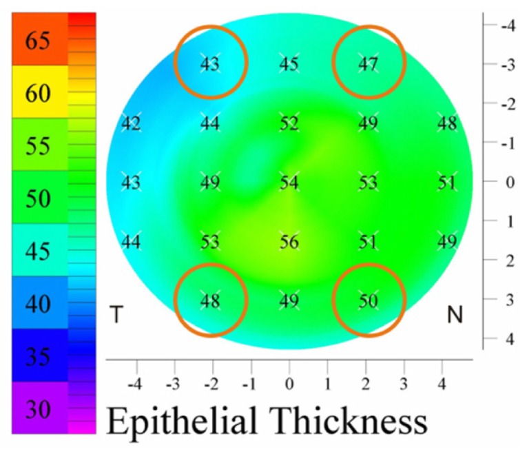
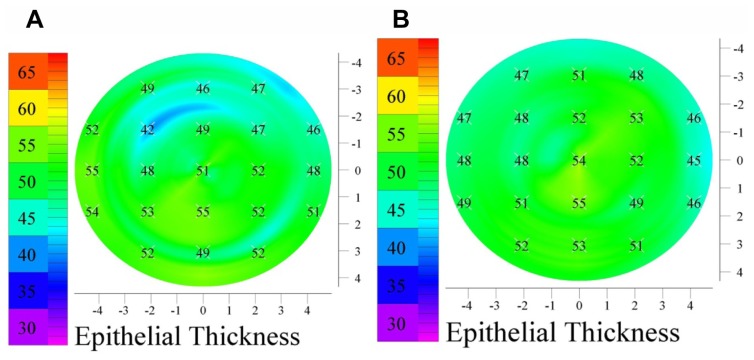
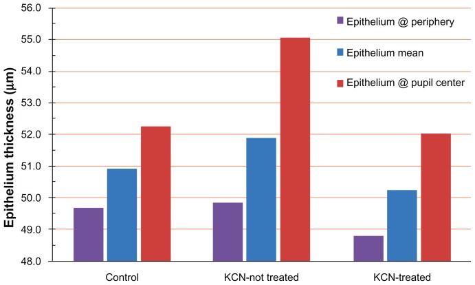
Epithelial pachymetry measurements (topographic mapping) were conducted on 100 subjects via HF UBM. Three groups of patients were included: patients with normal eyes (controls), patients with untreated keratoconic eyes, and patients with keratoconic eyes treated with CXL. Central, mean, and peripheral corneal epithelial thickness was examined for each group, and a statistical study was conducted.

RESULTS

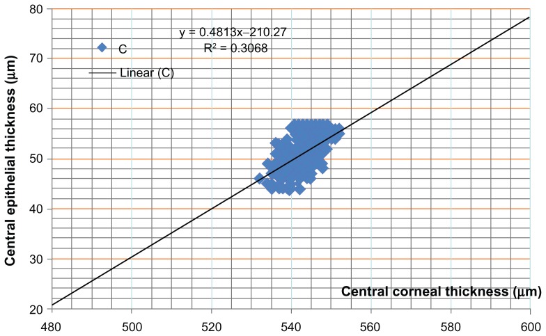
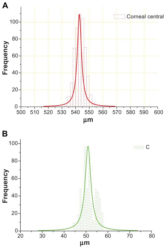
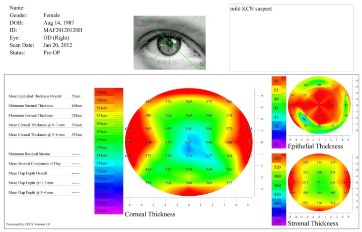
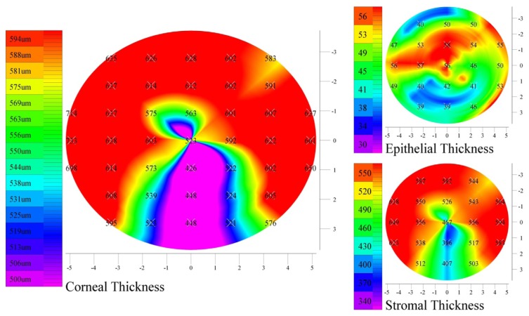
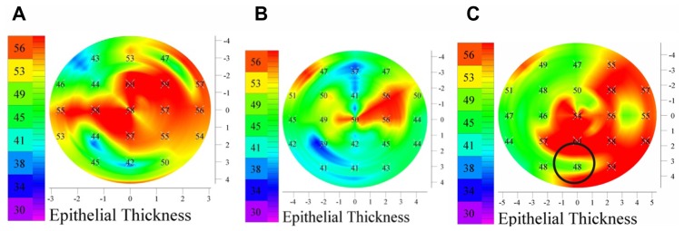
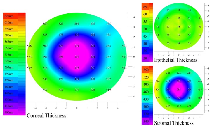
Mean, central, and peripheral corneal epithelial thickness was compared between the three groups of patients. Epithelium thickness varied substantially in the keratoconic group, and in some cases there was a difference of up to 20 μm between various points of the same eye, and often a thinner epithelium coincided with a thinner cornea. However, on average, data from the keratoconic group suggested an overall thickening of the epithelium, particularly over the pupil center of the order of +3 μm, while the mean epithelium thickness was on average +1.1 μm, compared to the control population (P = 0.005). This overall thickening was more pronounced in younger patients in the keratoconic group. Keratoconic eyes previously treated with CXL showed, on average, virtually the same average epithelium thickness (mean -0.7 μm, -0.2 μm over the pupil center, -0.9 μm over the peripheral zone) as the control group. This finding further reinforces our novel theory of the "reactive" component of epithelial thickening in corneas that are biomechanically unstable, becoming stable when biomechanical rigidity is accomplished despite persistence of cornea topographic irregularity.

CONCLUSION

A highly irregular epithelium may be suggestive of an ectatic cornea. Our results indicate that the epithelium is thinner over the keratoconic protrusion, but to a much lesser extent than anticipated, and on average epithelium is thicker in this group of patients. This difference appears to be clinically significant and may become a screening tool for eyes suspected for ectasia.

摘要

目的

使用数字弧形扫描超高频超声生物显微镜(HF UBM)成像系统(Artemis-II)测定角膜上皮厚度(测厚)并进行相关性分析,比较正常眼、未经治疗的圆锥角膜眼以及先前接受过胶原交联(CXL)治疗的圆锥角膜眼的平均上皮厚度和中央上皮厚度。

方法

通过HF UBM对100名受试者进行上皮测厚测量(地形图绘制)。纳入三组患者:正常眼患者(对照组)、未经治疗的圆锥角膜眼患者以及接受过CXL治疗的圆锥角膜眼患者。对每组的中央、平均和周边角膜上皮厚度进行检查,并进行统计学研究。

结果

比较了三组患者的平均、中央和周边角膜上皮厚度。圆锥角膜组的上皮厚度差异很大,在某些情况下,同一只眼的不同点之间相差可达20μm,而且上皮变薄往往与角膜变薄同时出现。然而,平均而言,圆锥角膜组的数据表明上皮总体增厚,尤其是在瞳孔中心上方约+3μm,而平均上皮厚度平均为+1.1μm,与对照组相比(P = 0.005)。这种总体增厚在圆锥角膜组的年轻患者中更为明显。先前接受过CXL治疗的圆锥角膜眼平均显示出与对照组几乎相同的平均上皮厚度(平均-0.7μm,瞳孔中心上方-0.2μm,周边区域-0.9μm)。这一发现进一步强化了我们关于生物力学不稳定角膜上皮增厚的“反应性”成分的新理论,即尽管角膜地形图不规则持续存在,但当生物力学刚度实现时,角膜上皮会变得稳定。

结论

高度不规则的上皮可能提示角膜扩张。我们的结果表明,在圆锥角膜突出部位上皮较薄,但程度远低于预期,并且该组患者的上皮平均更厚。这种差异似乎具有临床意义,可能成为疑似扩张眼的筛查工具。

https://cdn.ncbi.nlm.nih.gov/pmc/blobs/a57b/3373227/68dee14ee128/opth-6-789f12.jpg
https://cdn.ncbi.nlm.nih.gov/pmc/blobs/a57b/3373227/42e66a5920c5/opth-6-789f1.jpg
https://cdn.ncbi.nlm.nih.gov/pmc/blobs/a57b/3373227/b16f355e53c5/opth-6-789f2.jpg
https://cdn.ncbi.nlm.nih.gov/pmc/blobs/a57b/3373227/7cb60a67b79e/opth-6-789f3.jpg
https://cdn.ncbi.nlm.nih.gov/pmc/blobs/a57b/3373227/da9ad88b7379/opth-6-789f4.jpg
https://cdn.ncbi.nlm.nih.gov/pmc/blobs/a57b/3373227/e346c59896fd/opth-6-789f5.jpg
https://cdn.ncbi.nlm.nih.gov/pmc/blobs/a57b/3373227/28f96d9ec91f/opth-6-789f6.jpg
https://cdn.ncbi.nlm.nih.gov/pmc/blobs/a57b/3373227/f03794cd2636/opth-6-789f7.jpg
https://cdn.ncbi.nlm.nih.gov/pmc/blobs/a57b/3373227/7a0d779ade8c/opth-6-789f8.jpg
https://cdn.ncbi.nlm.nih.gov/pmc/blobs/a57b/3373227/ca08a0f1e477/opth-6-789f9.jpg
https://cdn.ncbi.nlm.nih.gov/pmc/blobs/a57b/3373227/e59c0b83f6d6/opth-6-789f10.jpg
https://cdn.ncbi.nlm.nih.gov/pmc/blobs/a57b/3373227/784aaff674e2/opth-6-789f11.jpg
https://cdn.ncbi.nlm.nih.gov/pmc/blobs/a57b/3373227/68dee14ee128/opth-6-789f12.jpg

相似文献

1
Correlation between epithelial thickness in normal corneas, untreated ectatic corneas, and ectatic corneas previously treated with CXL; is overall epithelial thickness a very early ectasia prognostic factor?正常角膜、未经治疗的扩张性角膜以及先前接受角膜交联治疗的扩张性角膜的上皮厚度之间的相关性;总体上皮厚度是否是早期扩张的预后因素?
Clin Ophthalmol. 2012;6:789-800. doi: 10.2147/OPTH.S31524. Epub 2012 May 23.
2
Meta-analysis of Pentacam vs. ultrasound pachymetry in central corneal thickness measurement in normal, post-LASIK or PRK, and keratoconic or keratoconus-suspect eyes.Pentacam 与超声角膜测厚仪在正常、LASIK 术后或 PRK 后、圆锥角膜或疑似圆锥角膜眼中中央角膜厚度测量的荟萃分析。
Graefes Arch Clin Exp Ophthalmol. 2014 Jan;252(1):91-9. doi: 10.1007/s00417-013-2502-5. Epub 2013 Nov 12.
3
Biomechanical assessment of healthy and keratoconic corneas (with/without crosslinking) using dynamic ultrahigh-speed Scheimpflug technology and the relevance of the parameter (A1L-A2L).利用动态超高速度 Scheimpflug 技术对健康角膜和圆锥角膜(交联/不交联)进行生物力学评估,以及参数(A1L-A2L)的相关性。
Br J Ophthalmol. 2019 Apr;103(4):558-564. doi: 10.1136/bjophthalmol-2017-311627. Epub 2018 Jun 5.
4
Comparison of efficacy and safety of accelerated trans-epithelial crosslinking for keratoconus patients with corneas thicker and thinner than 380μm.比较角膜厚度大于 380μm 和小于 380μm 的圆锥角膜患者行加速经上皮角膜交联术的疗效和安全性。
Curr Eye Res. 2022 Apr;47(4):511-516. doi: 10.1080/02713683.2021.2018466. Epub 2021 Dec 27.
5
Epithelial and stromal remodeling after corneal collagen cross-linking evaluated by spectral-domain OCT.应用频域光相干断层扫描评估角膜胶原交联术后上皮和基质重塑。
J Refract Surg. 2014 Feb;30(2):122-7. doi: 10.3928/1081597X-20140120-08.
6
Corneal epithelial thickness mapping using Fourier-domain optical coherence tomography for detection of form fruste keratoconus.使用傅里叶域光学相干断层扫描进行角膜上皮厚度测绘以检测forme fruste圆锥角膜。
J Cataract Refract Surg. 2015 Apr;41(4):812-20. doi: 10.1016/j.jcrs.2014.06.043.
7
Comparison of Corneal Epithelial and Stromal Thickness between Keratoconic and Normal Eyes in an Asian Population.亚洲人群中圆锥角膜与正常眼角膜上皮和基质厚度的比较。
Ophthalmic Res. 2019;62(3):134-140. doi: 10.1159/000500313. Epub 2019 Jul 2.
8
Evaluation of central and peripheral corneal thickness with ultrasound biomicroscopy in normal and keratoconic eyes.
Cornea. 1997 Nov;16(6):639-44.
9
SD-OCT analysis of regional epithelial thickness profiles in keratoconus, postoperative corneal ectasia, and normal eyes.SD-OCT 分析圆锥角膜、术后角膜扩张和正常眼中的区域性上皮厚度曲线。
J Refract Surg. 2013 Mar;29(3):173-9. doi: 10.3928/1081597X-20130129-08.
10
Corneal epithelial thickness mapping by Fourier-domain optical coherence tomography in normal and keratoconic eyes.傅里叶域光学相干断层扫描对正常眼和圆锥角膜眼的角膜上皮厚度测绘。
Ophthalmology. 2012 Dec;119(12):2425-33. doi: 10.1016/j.ophtha.2012.06.023. Epub 2012 Aug 20.

引用本文的文献

1
Corneal epithelium is altered in keratoconus and forme fruste keratoconus.圆锥角膜和亚临床圆锥角膜的角膜上皮会发生改变。
Sci Rep. 2025 Jul 1;15(1):22203. doi: 10.1038/s41598-025-07663-6.
2
Corneal Epithelial Thickness Maps in Eyes with Mild and Moderate Keratoconus.轻度和中度圆锥角膜患者的角膜上皮厚度图
J Clin Med. 2025 Feb 14;14(4):1256. doi: 10.3390/jcm14041256.
3
Corneal Epithelial Thickness Mapping in Healthy Population Corneas Using MS-39 Anterior Segment Optical Coherence Tomography.使用MS-39眼前节光学相干断层扫描技术对健康人群角膜进行角膜上皮厚度测绘

本文引用的文献

1
Corneal epithelial thickness mapping by Fourier-domain optical coherence tomography in normal and keratoconic eyes.傅里叶域光学相干断层扫描对正常眼和圆锥角膜眼的角膜上皮厚度测绘。
Ophthalmology. 2012 Dec;119(12):2425-33. doi: 10.1016/j.ophtha.2012.06.023. Epub 2012 Aug 20.
2
Long term results of a prospective randomized bilateral eye comparison trial of higher fluence, shorter duration ultraviolet A radiation, and riboflavin collagen cross linking for progressive keratoconus.一项关于高能量、短持续时间紫外线A辐射联合核黄素胶原交联治疗进行性圆锥角膜的前瞻性随机双眼对照试验的长期结果
Clin Ophthalmol. 2012;6:97-101. doi: 10.2147/OPTH.S27170. Epub 2012 Jan 11.
3
Clin Ophthalmol. 2025 Jan 22;19:249-259. doi: 10.2147/OPTH.S503195. eCollection 2025.
4
The Corneal Epithelial Thickness Profile in a Healthy Saudi Population.沙特健康人群的角膜上皮厚度分布
Cureus. 2024 Oct 9;16(10):e71135. doi: 10.7759/cureus.71135. eCollection 2024 Oct.
5
Repeatability of Corneal Epithelial Thickness Map Using Anterior-Segment Optical Coherence Tomography in Normal and Corneal Disease Patients.眼前节光学相干断层扫描测量正常人和角膜病患者角膜上皮厚度图的可重复性
Korean J Ophthalmol. 2024 Dec;38(6):496-506. doi: 10.3341/kjo.2024.0115. Epub 2024 Oct 22.
6
Topographic Keratoconus Incidence in Greece Diagnosed in Routine Consecutive Cataract Procedures: A Consecutive Case Series of 1250 Cases over 5 Years.希腊常规连续性白内障手术中诊断出的圆锥角膜发病率:一项5年期间1250例连续病例系列研究。
J Clin Med. 2024 Apr 19;13(8):2378. doi: 10.3390/jcm13082378.
7
Keratoconus: exploring fundamentals and future perspectives - a comprehensive systematic review.圆锥角膜:探索基础与未来展望——一项全面的系统综述
Ther Adv Ophthalmol. 2024 Mar 20;16:25158414241232258. doi: 10.1177/25158414241232258. eCollection 2024 Jan-Dec.
8
Ray-Tracing Customization in Myopic and Myopic Astigmatism LASIK Treatments for Low and High Order Aberrations Treatment: 2-Year Visual Function and Psychometric Value Outcomes of a Consecutive Case Series.近视及近视散光LASIK手术中用于低阶和高阶像差治疗的光线追踪定制:连续病例系列的2年视觉功能和心理测量值结果
Clin Ophthalmol. 2024 Feb 22;18:565-574. doi: 10.2147/OPTH.S444174. eCollection 2024.
9
Corneal Epithelial Thickness Mapping: A Major Review.角膜上皮厚度地形图:一项主要综述。
J Ophthalmol. 2024 Jan 2;2024:6674747. doi: 10.1155/2024/6674747. eCollection 2024.
10
Assessing progression limits in different grades of keratoconus from a novel perspective: precision of measurements of the corneal epithelium.从新视角评估不同等级圆锥角膜的进展限度:角膜上皮测量的精确性
Eye Vis (Lond). 2024 Jan 2;11(1):1. doi: 10.1186/s40662-023-00368-9.
Corneal responses to eye rubbing with spectral domain optical coherence tomography.
利用频域光学相干断层扫描技术观察眼球摩擦对角膜的反应。
Curr Eye Res. 2012 Jan;37(1):25-32. doi: 10.3109/02713683.2011.622850. Epub 2011 Oct 26.
4
Comparison of Scheimpflug imaging and spectral domain anterior segment optical coherence tomography for detection of narrow anterior chamber angles.Scheimpflug 成像与频域眼前节光学相干断层扫描检测窄房角的比较。
Eye (Lond). 2011 May;25(5):603-11. doi: 10.1038/eye.2011.14. Epub 2011 Feb 18.
5
Comparison of sequential vs same-day simultaneous collagen cross-linking and topography-guided PRK for treatment of keratoconus.序贯式与同日同步胶原交联联合地形图引导的准分子激光原位角膜磨镶术治疗圆锥角膜的比较
J Refract Surg. 2009 Sep;25(9):S812-8. doi: 10.3928/1081597X-20090813-10. Epub 2009 Sep 11.
6
Corneal endothelium in patients with diabetes mellitus: a historical cohort study.糖尿病患者的角膜内皮:一项历史性队列研究。
Eur J Ophthalmol. 2009 May-Jun;19(3):369-75. doi: 10.1177/112067210901900307.
7
Epithelial thickness in the normal cornea: three-dimensional display with Artemis very high-frequency digital ultrasound.正常角膜的上皮厚度:使用阿耳忒弥斯超高频率数字超声进行三维显示。
J Refract Surg. 2008 Jun;24(6):571-81. doi: 10.3928/1081597X-20080601-05.
8
Very-high-frequency ultrasound corneal imaging as a new tool for early diagnosis of ocular surface toxicity in rabbits treated with a preserved glaucoma drug.甚高频超声角膜成像作为一种新工具用于早期诊断用一种保存型青光眼药物治疗的家兔眼表毒性。
Ophthalmic Res. 2008 Oct;40(6):298-308. doi: 10.1159/000134928. Epub 2008 May 28.
9
The repeatability of corneal and corneal epithelial thickness measurements using optical coherence tomography.使用光学相干断层扫描技术测量角膜及角膜上皮厚度的可重复性。
Optom Vis Sci. 2006 Jun;83(6):360-5. doi: 10.1097/01.opx.0000221388.26031.23.
10
Corneal and epithelial thickness in keratoconus: a comparison of ultrasonic pachymetry, Orbscan II, and optical coherence tomography.圆锥角膜的角膜和上皮厚度:超声测厚仪、Orbscan II和光学相干断层扫描的比较
J Refract Surg. 2006 May;22(5):486-93. doi: 10.3928/1081-597X-20060501-11.