• 文献检索
  • 文档翻译
  • 深度研究
  • 学术资讯
  • Suppr Zotero 插件Zotero 插件
  • 邀请有礼
  • 套餐&价格
  • 历史记录
应用&插件
Suppr Zotero 插件Zotero 插件浏览器插件Mac 客户端Windows 客户端微信小程序
定价
高级版会员购买积分包购买API积分包
服务
文献检索文档翻译深度研究API 文档MCP 服务
关于我们
关于 Suppr公司介绍联系我们用户协议隐私条款
关注我们

Suppr 超能文献

核心技术专利:CN118964589B侵权必究
粤ICP备2023148730 号-1Suppr @ 2026

文献检索

告别复杂PubMed语法,用中文像聊天一样搜索,搜遍4000万医学文献。AI智能推荐,让科研检索更轻松。

立即免费搜索

文件翻译

保留排版,准确专业,支持PDF/Word/PPT等文件格式,支持 12+语言互译。

免费翻译文档

深度研究

AI帮你快速写综述,25分钟生成高质量综述,智能提取关键信息,辅助科研写作。

立即免费体验

蛙眼单个视杆外段的 X 射线衍射研究。

An X-ray diffraction study on a single rod outer segment from frog retina.

机构信息

Japan Synchrotron Radiation Research Institute, SPring-8, 1-1-1 Kouto, Sayo-cho, Sayo-gun, Hyogo 679-5198, Japan.

出版信息

J Synchrotron Radiat. 2012 Jul;19(Pt 4):574-8. doi: 10.1107/S0909049512018535. Epub 2012 May 18.

DOI:10.1107/S0909049512018535
PMID:22713892
原文链接:https://pmc.ncbi.nlm.nih.gov/articles/PMC3380658/
Abstract

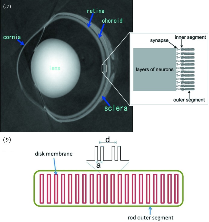
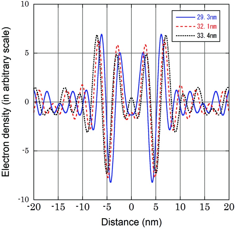
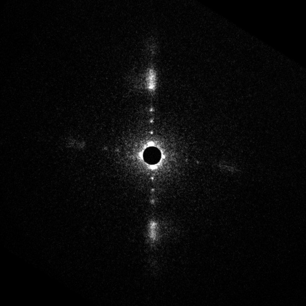
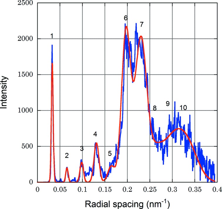
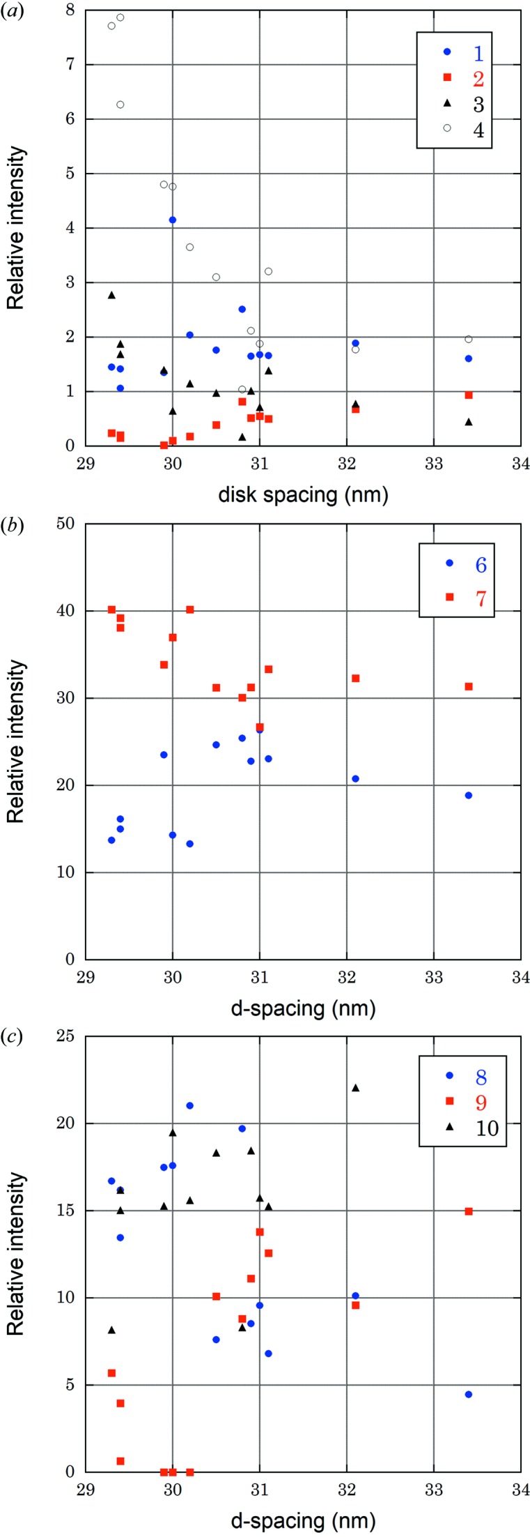
X-ray diffraction patterns were recorded from isolated single rod outer segments of frog. The outer segments in Ringer's solution were exposed to a 6 µm microbeam (15 keV) at the BL40XU beamline, SPring-8. The diffraction pattern demonstrated a remarkable regularity in the stacking and flatness of the disk membranes. The electron density profile calculated from the intensity of up to tenth-order reflections showed a pair of bilayers that comprise a disk membrane. The structure of the disk membrane and the changes in the profile on swelling generally agreed with previous reports. Radiation damage was significant with an irradiation of 5 × 10(5) Gy which is much lower than the known damaging dose on proteins at the liquid-nitrogen temperature.

摘要

我们从青蛙的分离的单个杆状部的外节段中记录了 X 射线衍射图谱。在 Ringer 溶液中的外节段在 SPring-8 的 BL40XU 光束线上暴露于 6μm 的微光束(15keV)下。衍射图谱显示出盘膜的堆叠和平整度的显著规律性。从高达第十阶反射强度计算出的电子密度分布显示出一对双层,其构成了盘膜。盘膜的结构和肿胀时轮廓的变化通常与以前的报告一致。辐照损伤非常显著,辐照剂量为 5×10(5)Gy,远低于在液氮温度下已知的蛋白质损伤剂量。

https://cdn.ncbi.nlm.nih.gov/pmc/blobs/60a7/3380658/13955210e375/s-19-00574-fig5.jpg
https://cdn.ncbi.nlm.nih.gov/pmc/blobs/60a7/3380658/2bf89d2f332b/s-19-00574-fig1.jpg
https://cdn.ncbi.nlm.nih.gov/pmc/blobs/60a7/3380658/7e555ebfb8a9/s-19-00574-fig2.jpg
https://cdn.ncbi.nlm.nih.gov/pmc/blobs/60a7/3380658/7534e69ee374/s-19-00574-fig3.jpg
https://cdn.ncbi.nlm.nih.gov/pmc/blobs/60a7/3380658/3dcfb8737137/s-19-00574-fig4.jpg
https://cdn.ncbi.nlm.nih.gov/pmc/blobs/60a7/3380658/13955210e375/s-19-00574-fig5.jpg

相似文献

1
An X-ray diffraction study on a single rod outer segment from frog retina.蛙眼单个视杆外段的 X 射线衍射研究。
J Synchrotron Radiat. 2012 Jul;19(Pt 4):574-8. doi: 10.1107/S0909049512018535. Epub 2012 May 18.
2
A 12 A resolution X-ray diffraction study of the profile structure of isolated bovine retinal rod outer segment disk membranes.对分离的牛视网膜杆状外段盘膜轮廓结构的12A分辨率X射线衍射研究。
Biochim Biophys Acta. 1984 Oct 17;777(1):9-20. doi: 10.1016/0005-2736(84)90491-7.
3
[Molecular mechanisms of photoreception. V. Isolation of cytoplasmic membranes of rod outer segments from the frog retina].[光感受器的分子机制。V. 从青蛙视网膜分离视杆细胞外段的细胞质膜]
Mol Biol (Mosk). 1986 Nov-Dec;20(6):1479-87.
4
Evaluation of the electron density profile of the frog rod outer segment disc-membrane in vivo using x-ray diffraction.利用X射线衍射对青蛙视杆细胞外段盘膜的电子密度分布进行体内评估。
Biochim Biophys Acta. 1981 Jan 8;640(1):142-58. doi: 10.1016/0005-2736(81)90540-x.
5
Localization of peripherin/rds in the disk membranes of cone and rod photoreceptors: relationship to disk membrane morphogenesis and retinal degeneration.外周蛋白/视网膜变性慢病毒(peripherin/rds)在视锥和视杆光感受器盘膜中的定位:与盘膜形态发生和视网膜变性的关系。
J Cell Biol. 1992 Feb;116(3):659-67. doi: 10.1083/jcb.116.3.659.
6
The role of taurine in osmotic, mechanical, and chemical protection of the retinal rod outer segments.牛磺酸在视网膜视杆细胞外段的渗透、机械和化学保护中的作用。
Adv Exp Med Biol. 1998;442:407-13. doi: 10.1007/978-1-4899-0117-0_50.
7
X-ray and neutron diffraction of retinal rod outer segments.视网膜杆状细胞外段的X射线和中子衍射
Methods Enzymol. 1982;81:593-604. doi: 10.1016/s0076-6879(82)81083-5.
8
X-ray diffraction and electron microscope study of phase separation in rod outer segment photoreceptor membrane multilayers.杆状外段光感受器膜多层结构中相分离的X射线衍射和电子显微镜研究。
Biophys J. 1982 Sep;39(3):241-51. doi: 10.1016/S0006-3495(82)84514-1.
9
Sialoglycoproteins of the frog rod outer segment plasma membrane.青蛙视杆细胞外节质膜的唾液酸糖蛋白。
Invest Ophthalmol Vis Sci. 1988 Oct;29(10):1523-32.
10
Na+- and cGMP-induced Ca2+ fluxes in frog rod photoreceptors.蛙类视杆光感受器中钠离子和环鸟苷酸诱导的钙离子通量。
J Gen Physiol. 1987 Mar;89(3):481-500. doi: 10.1085/jgp.89.3.481.

引用本文的文献

1
Fiber Diffraction and Small-Angle Scattering for Structural Investigation of Bacterial Amyloids.用于细菌淀粉样蛋白结构研究的纤维衍射和小角散射
Methods Mol Biol. 2022;2538:95-107. doi: 10.1007/978-1-0716-2529-3_7.

本文引用的文献

1
Optical properties of in situ eye lenses measured with X-ray Talbot interferometry: a novel measure of growth processes.用 X 射线泰伯干涉法测量原位晶状体的光学特性:一种测量生长过程的新方法。
PLoS One. 2011;6(9):e25140. doi: 10.1371/journal.pone.0025140. Epub 2011 Sep 20.
2
The partition of cholesterol between ordered and fluid bilayers of phosphatidylcholine: a synchrotron X-ray diffraction study.胆固醇在磷脂酰胆碱有序双层和流体双层之间的分配:一项同步加速器X射线衍射研究。
Biochim Biophys Acta. 2007 Nov;1768(11):2873-81. doi: 10.1016/j.bbamem.2007.07.023. Epub 2007 Aug 16.
3
Three-dimensional architecture of murine rod outer segments determined by cryoelectron tomography.
通过冷冻电子断层扫描确定的小鼠视杆细胞外段的三维结构
J Cell Biol. 2007 Jun 4;177(5):917-25. doi: 10.1083/jcb.200612010. Epub 2007 May 29.
4
An x-ray diffraction study on early structural changes in skeletal muscle contraction.一项关于骨骼肌收缩早期结构变化的X射线衍射研究。
Biophys J. 2003 Feb;84(2 Pt 1):1093-102. doi: 10.1016/S0006-3495(03)74925-X.
5
Potential for biomolecular imaging with femtosecond X-ray pulses.飞秒X射线脉冲用于生物分子成像的潜力。
Nature. 2000 Aug 17;406(6797):752-7. doi: 10.1038/35021099.
6
Neutron diffraction analysis of the structure of rod photoreceptor membranes in intact retinas.完整视网膜中视杆光感受器膜结构的中子衍射分析
J Mol Biol. 1980 Mar 5;137(3):315-48. doi: 10.1016/0022-2836(80)90319-8.
7
Structure of frog photoreceptor membranes.青蛙光感受器膜的结构。
Nature. 1969 Aug 30;223(5209):906-9. doi: 10.1038/223906a0.
8
Molecular localization of frog retinal receptor photopigment by electron microscopy and low-angle X-ray diffraction.通过电子显微镜和低角度X射线衍射对青蛙视网膜受体光色素进行分子定位
J Mol Biol. 1969 Feb 14;39(3):407-16. doi: 10.1016/0022-2836(69)90135-1.
9
X-ray analysis of retinal photoreceptors.视网膜光感受器的X射线分析。
Proc Natl Acad Sci U S A. 1969 Jun;63(2):233-8. doi: 10.1073/pnas.63.2.233.
10
Lamellar structure of bleached and unbleached rod photoreceptor membranes.漂白和未漂白的视杆光感受器膜的层状结构。
Nature. 1972 May 26;237(5352):229-31. doi: 10.1038/237229a0.