University Eye Hospital Freiburg, Freiburg, Germany.
PLoS One. 2012;7(6):e38820. doi: 10.1371/journal.pone.0038820. Epub 2012 Jun 18.
Disturbed axonal transport is an important pathogenic factor in many neurodegenerative diseases, such as glaucoma, an eye disease characterised by progressive atrophy of the optic nerve. Quantification of retrograde axonal transport in the optic nerve usually requires labour intensive histochemical techniques or expensive equipment for in vivo imaging. Here, we report on a robust alternative method using Fluorogold (FG) as tracer, which is spectrometrically quantified in retinal tissue lysate.
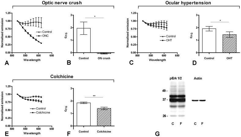
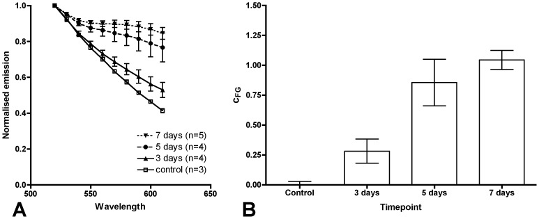
To determine parameters reflecting the relative FG content of a sample FG was dissolved in retinal lysates at different concentrations and spectra were obtained. For validation in vivo FG was injected uni- or bilaterally into the superior colliculus (SC) of Sprague Dawley rats. The retinal lysate was analysed after 3, 5 and 7 days to determine the time course of FG accumulation in the retina (n = 15). In subsequent experiments axona transport was impaired by optic nerve crush (n = 3), laser-induced ocular hypertension (n = 5) or colchicine treatment to the SC (n = 10).
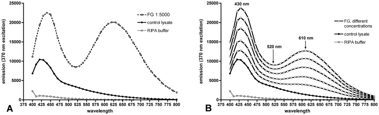
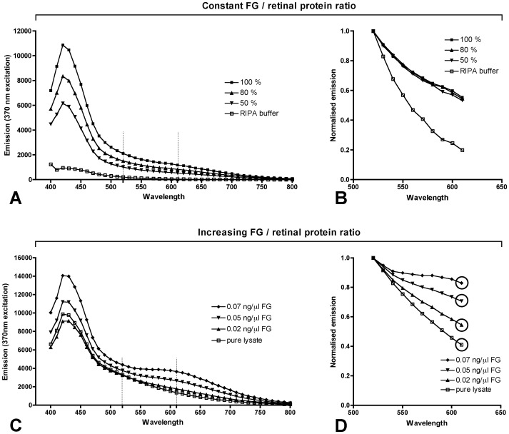
Spectrometry at 370 nm excitation revealed two emission peaks at 430 and 610 nm. We devised a formula to calculate the relative FG content (c(FG)), from the emission spectrum. c(FG) is proportional to the real FG concentration as it corrects for variations of retinal protein concentration in the lysate. After SC injection, c(FG) monotonously increases with time (p = 0.002). Optic nerve axonal damage caused a significant decrease of c(FG) (crush p = 0.029; hypertension p = 0.025; colchicine p = 0.006). Lysates are amenable to subsequent protein analysis.
Spectrometrical FG detection in retinal lysates allows for quantitative assessment of retrograde axonal transport using standard laboratory equipment. It is faster than histochemical techniques and may also complement morphological in vivo analyses.
轴突运输障碍是许多神经退行性疾病的重要致病因素,例如青光眼,这是一种以视神经进行性萎缩为特征的眼部疾病。视神经逆行轴突运输的定量通常需要费力的组织化学技术或昂贵的体内成像设备。在这里,我们报告了一种使用荧光金(FG)作为示踪剂的强大替代方法,该方法可通过光谱法在视网膜组织裂解物中进行定量。
为了确定反映样品中 FG 相对含量的参数,将 FG 溶解在视网膜裂解物中的不同浓度下,并获得光谱。为了进行体内验证,将 FG 单侧或双侧注射到 Sprague Dawley 大鼠的上丘(SC)中。在第 3、5 和 7 天分析视网膜裂解物,以确定 FG 在视网膜中的积累时间过程(n = 15)。在随后的实验中,通过视神经挤压(n = 3)、激光诱导的眼高压(n = 5)或 SC 中的秋水仙碱处理(n = 10)来损害轴突运输。
在 370nm 激发下的光谱法显示出两个发射峰,分别在 430nm 和 610nm 处。我们设计了一个公式,从发射光谱中计算出相对 FG 含量(c(FG))。c(FG)与真实 FG 浓度成正比,因为它校正了裂解物中视网膜蛋白浓度的变化。SC 注射后,c(FG)随时间单调增加(p = 0.002)。视神经轴突损伤导致 c(FG)显著降低(挤压 p = 0.029;高血压 p = 0.025;秋水仙碱 p = 0.006)。裂解物适用于后续的蛋白质分析。
视网膜裂解物中荧光金的光谱检测允许使用标准实验室设备对逆行轴突运输进行定量评估。它比组织化学技术更快,也可能补充体内形态学分析。