Department of Neuroscience and Imaging, Institute for Advanced Biomedical Technologies (ITAB), University G. d'Annunzio of Chieti, Chieti, Italy.
PLoS One. 2012;7(6):e39118. doi: 10.1371/journal.pone.0039118. Epub 2012 Jun 18.
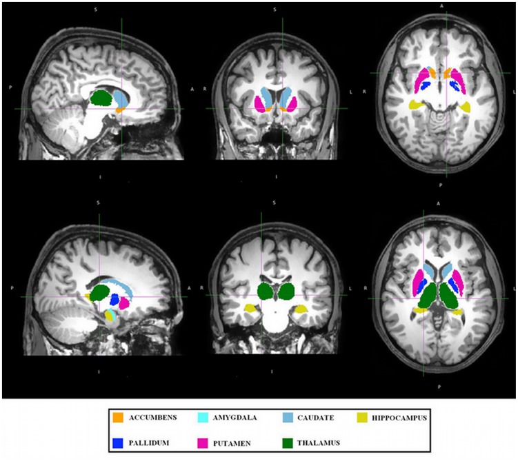
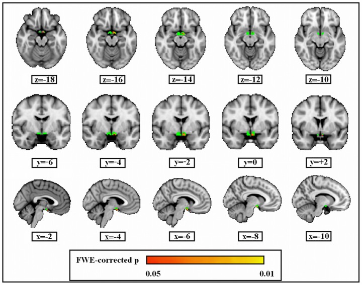
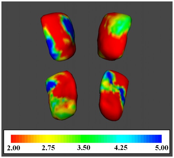
Psychogenic erectile dysfunction (ED) has been defined as the persistent inability to attain and maintain an erection sufficient to permit sexual performance. It shows a high incidence and prevalence among men, with a significant impact on the quality of life. Few neuroimaging studies have investigated the cerebral basis of erectile dysfunctions observing the role played by prefrontal, cingulate, and parietal cortices during erotic stimulation. In spite of the well-known involvement of subcortical regions such as hypothalamus and caudate nucleus in male sexual response, and the key role of nucleus accumbens in pleasure and reward, poor attention was paid to their role in male sexual dysfunction. In this study, we determined the presence of grey matter (GM) atrophy patterns in subcortical structures such as amygdala, hippocampus, nucleus accumbens, caudate nucleus, putamen, pallidum, thalamus, and hypothalamus in patients with psychogenic ED and healthy men. After Rigiscan evaluation, urological, general medical, metabolic and hormonal, psychological and psychiatric assessment, 17 outpatients with psychogenic ED and 25 healthy controls were recruited for structural MRI session. Significant GM atrophy of nucleus accumbens was observed bilaterally in patients with respect to controls. Shape analysis showed that this atrophy was located in the left medial-anterior and posterior portion of accumbens. Left nucleus accumbens volumes in patients correlated with low erectile functioning as measured by IIEF-5 (International Index of Erectile Function). In addition, a GM atrophy of left hypothalamus was also observed. Our results suggest that atrophy of nucleus accumbens plays an important role in psychogenic erectile dysfunction. We believe that this change can influence the motivation-related component of sexual behavior. Our findings help to elucidate a neural basis of psychogenic erectile dysfunction.
心因性勃起功能障碍(ED)被定义为持续无法获得和维持足以进行性行为的勃起。它在男性中发病率和患病率都很高,对生活质量有重大影响。少数神经影像学研究调查了勃起功能障碍的大脑基础,观察了在性刺激期间前额叶、扣带回和顶叶皮质的作用。尽管众所周知下丘脑和尾状核等皮质下区域在男性性反应中起重要作用,而伏隔核在愉悦和奖励中起关键作用,但人们对它们在男性性功能障碍中的作用关注甚少。在这项研究中,我们确定了心因性 ED 患者和健康男性的皮质下结构(如杏仁核、海马体、伏隔核、尾状核、壳核、苍白球、丘脑和下丘脑)中的灰质(GM)萎缩模式。在进行 Rigiscan 评估、泌尿科、一般医学、代谢和激素、心理和精神病评估后,招募了 17 名心因性 ED 门诊患者和 25 名健康对照者进行结构 MRI 检查。与对照组相比,心因性 ED 患者双侧伏隔核 GM 萎缩明显。形态分析显示,这种萎缩位于伏隔核的左侧内侧-前和后部分。患者的左侧伏隔核体积与 IIEF-5(国际勃起功能指数)测量的低勃起功能相关。此外,还观察到左侧下丘脑的 GM 萎缩。我们的研究结果表明,伏隔核的萎缩在心因性勃起功能障碍中起重要作用。我们认为这种变化可以影响性行为的动机相关成分。我们的研究结果有助于阐明心因性勃起功能障碍的神经基础。