Blizard Institute, Barts and The London School of Medicine and Dentistry, Queen Mary University of London, Core Pathology Facility, The Royal London Hospital, 80 Newark Street, London E1 2ES, UK.
Br J Cancer. 2012 Jul 24;107(3):477-81. doi: 10.1038/bjc.2012.268. Epub 2012 Jun 26.
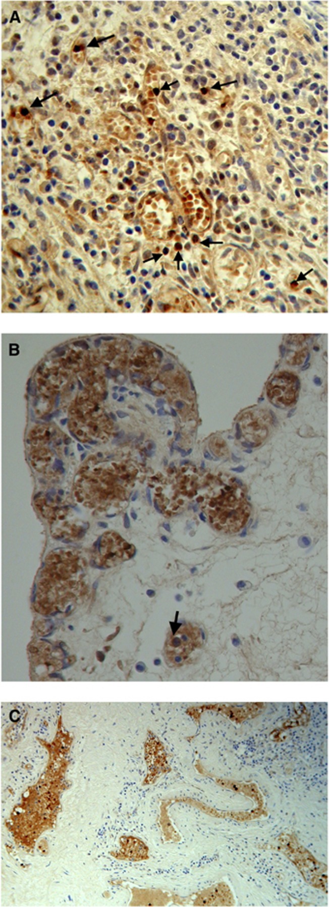
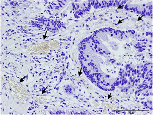
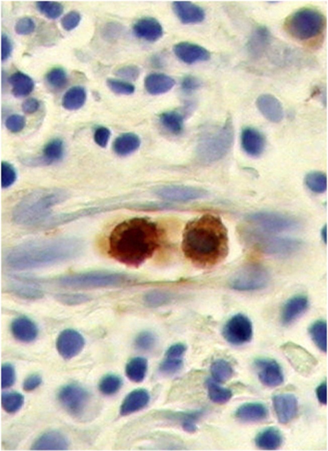
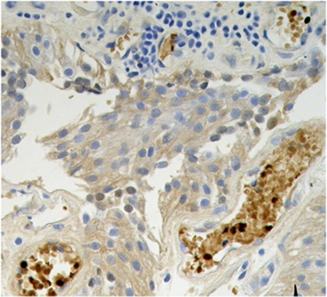
The immunohistochemical features of fetal haemoglobin cells and their distribution patterns in solid tumours, such as colorectal cancer and blastomas, suggest that fetal haemopoiesis may take place in these tumour tissues. These locally highly concentrated fetal haemoglobin (HbF) cells may promote tumour growth by providing a more efficient oxygen supply.
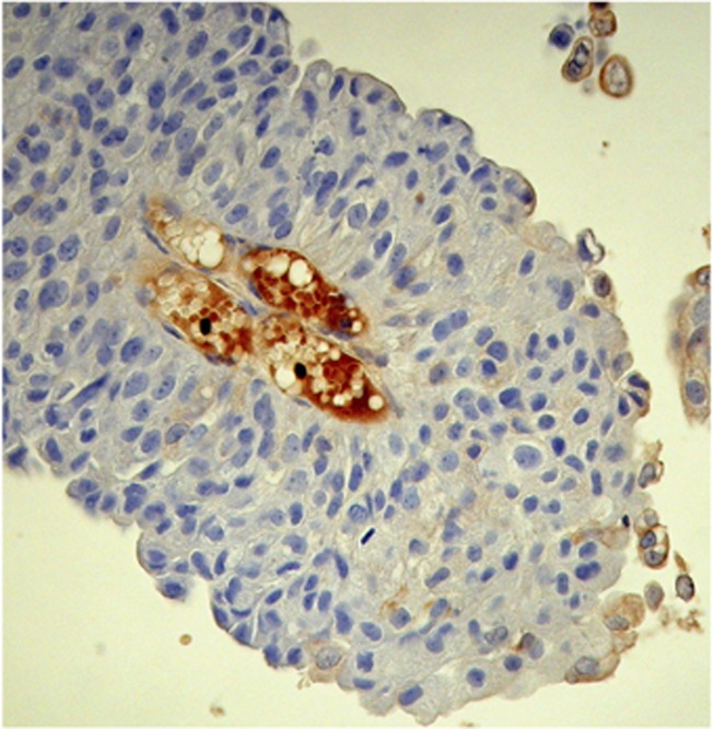
Biomarkers of HbF were checked in transitional cell carcinoma (TCC) of the urinary bladder, assessing this as a new parameter for disease management. Fetal haemoglobin was immunohistochemically examined in tumours from 60 patients with TCC of the bladder. Fetal haemoglobin erythrocytes and erythroblasts were mainly clonally distributed in proliferating blood vessels and not mixed with normal haemoglobin erythrocytes. The proportion of such HbF blood vessels could reach more than half of the total number of vessels. There were often many HbF erythroblasts distributed in one-cell or two-cell capillaries and present as 5-15% of cells in multi-cell vessels. This suggests a local proliferation of HbF-cell progenitors. Fetal haemoglobin cells were prominently marking lower grades of tumours, as 76% (n=21) of the patients with G1pTa were HbF+, whereas only 6.7% (n=30) of the patients with G3pT1-pT2a were HbF+.
Our results suggest that HbF, besides being a potential new marker for early tumour detection, might be an essential factor of early tumour development, as in fetal life. Inhibiting HbF upregulation may provide a therapeutic target for the inhibition of tumour growth.
胎儿血红蛋白细胞的免疫组织化学特征及其在结直肠癌和胚胎瘤等实体瘤中的分布模式表明,胎儿造血可能发生在这些肿瘤组织中。这些局部高度集中的胎儿血红蛋白 (HbF) 细胞可能通过提供更有效的氧气供应来促进肿瘤生长。
在膀胱癌中转移细胞癌 (TCC) 中检查了 HbF 的生物标志物,将其评估为疾病管理的新参数。在 60 例膀胱癌 TCC 肿瘤中免疫组织化学检查了胎儿血红蛋白。胎儿血红蛋白红细胞和幼红细胞主要在增殖的血管中呈克隆性分布,与正常血红蛋白红细胞不混合。这种 HbF 血管的比例可达到总血管数的一半以上。在一个或两个细胞的毛细血管中经常分布有许多 HbF 幼红细胞,并且在多细胞血管中占 5-15%的细胞。这表明 HbF 细胞祖细胞存在局部增殖。胎儿血红蛋白细胞明显标记肿瘤的低级别,因为 76%(n=21)的 G1pTa 患者为 HbF+,而只有 6.7%(n=30)的 G3pT1-pT2a 患者为 HbF+。
我们的结果表明,HbF 除了作为早期肿瘤检测的潜在新标志物外,可能是早期肿瘤发展的重要因素,就像在胎儿生命中一样。抑制 HbF 上调可能为抑制肿瘤生长提供治疗靶点。