• 文献检索
  • 文档翻译
  • 深度研究
  • 学术资讯
  • Suppr Zotero 插件Zotero 插件
  • 邀请有礼
  • 套餐&价格
  • 历史记录
应用&插件
Suppr Zotero 插件Zotero 插件浏览器插件Mac 客户端Windows 客户端微信小程序
定价
高级版会员购买积分包购买API积分包
服务
文献检索文档翻译深度研究API 文档MCP 服务
关于我们
关于 Suppr公司介绍联系我们用户协议隐私条款
关注我们

Suppr 超能文献

核心技术专利:CN118964589B侵权必究
粤ICP备2023148730 号-1Suppr @ 2026

文献检索

告别复杂PubMed语法,用中文像聊天一样搜索,搜遍4000万医学文献。AI智能推荐,让科研检索更轻松。

立即免费搜索

文件翻译

保留排版,准确专业,支持PDF/Word/PPT等文件格式,支持 12+语言互译。

免费翻译文档

深度研究

AI帮你快速写综述,25分钟生成高质量综述,智能提取关键信息,辅助科研写作。

立即免费体验

胃小黏膜下肿瘤:CT 对胃神经鞘瘤与胃肠道间质瘤的鉴别诊断。

Small submucosal tumors of the stomach: differentiation of gastric schwannoma from gastrointestinal stromal tumor with CT.

机构信息

Department of Radiology, Samsung Medical Center, Sungkyunkwan University School of Medicine, Seoul 135-710, Korea.

出版信息

Korean J Radiol. 2012 Jul-Aug;13(4):425-33. doi: 10.3348/kjr.2012.13.4.425. Epub 2012 Jun 18.

DOI:10.3348/kjr.2012.13.4.425
PMID:22778564
原文链接:https://pmc.ncbi.nlm.nih.gov/articles/PMC3384824/
Abstract

OBJECTIVE

To identify the CT features that help differentiate gastric schwannomas (GS) from small (5 cm or smaller) gastrointestinal stromal tumors (GIST) and to assess the growth rates of both tumors.

MATERIALS AND METHODS

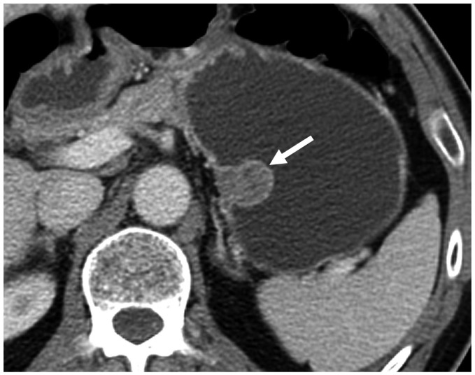
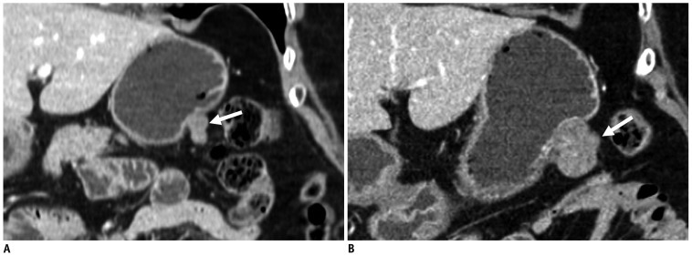
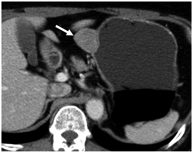
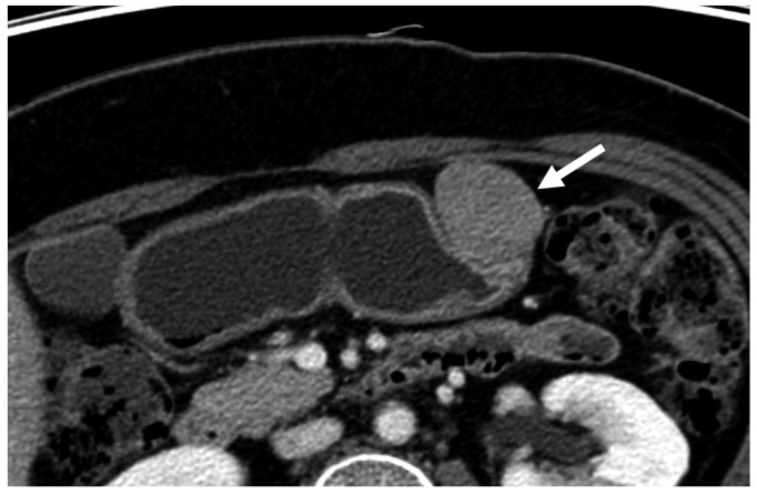
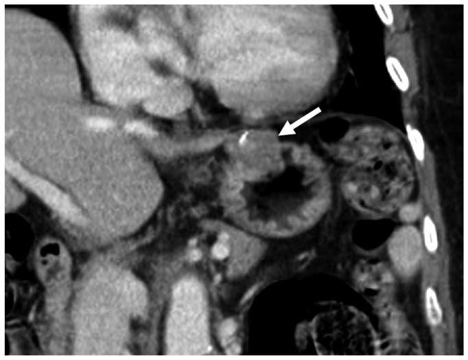
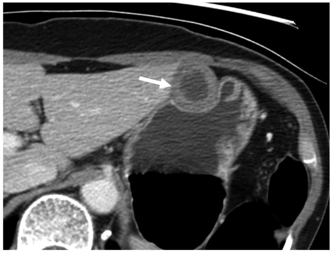
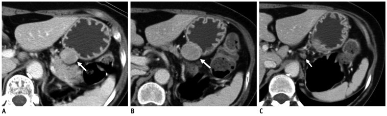
We included 16 small GSs and 56 GISTs located in the stomach. We evaluated the CT features including size, contour, surface pattern, margins, growth pattern, pattern and degree of contrast enhancement, and the presence of intralesional low attenuation area, hemorrhage, calcification, surface dimpling, fistula, perilesional lymph nodes (LNs), invasion to other organs, metastasis, ascites, and peritoneal seeding. We also estimated the tumor volume doubling time.

RESULTS

Compared with GISTs, GSs more frequently demonstrated a homogeneous enhancement pattern, exophytic or mixed growth pattern, and the presence of perilesional LNs (each p < 0.05). The intralesional low attenuation area was more common in GISTs than GSs (p < 0.05). Multivariate analyses indicated that a homogeneous enhancement pattern, exophytic or mixed growth pattern, and the presence of perilesional LNs were statistically significant (p < 0.05). Tumor volume doubling times for GSs (mean, 1685.4 days) were significantly longer than that of GISTs (mean, 377.6 days) (p = 0.004).

CONCLUSION

Although small GSs and GISTs show similar imaging findings, GSs more frequently show an exophytic or mixed growth pattern, homogeneous enhancement pattern, perilesional LNs and grow slower than GISTs.

摘要

目的

确定有助于区分胃 schwannomas(GS)和小(5cm 或更小)胃肠道间质瘤(GIST)的 CT 特征,并评估两种肿瘤的生长速度。

材料和方法

我们纳入了 16 个位于胃中的小 GS 和 56 个 GIST。我们评估了 CT 特征,包括大小、轮廓、表面形态、边缘、生长方式、对比增强模式和程度,以及内部低衰减区、出血、钙化、表面凹陷、瘘管、周围淋巴结(LN)、侵犯其他器官、转移、腹水和腹膜播种的存在。我们还估计了肿瘤体积倍增时间。

结果

与 GIST 相比,GS 更常表现出均匀强化模式、外生性或混合生长模式以及周围 LN 的存在(均 p<0.05)。内部低衰减区在 GIST 中比 GS 更常见(p<0.05)。多变量分析表明,均匀强化模式、外生性或混合生长模式以及周围 LN 的存在具有统计学意义(p<0.05)。GS 的肿瘤体积倍增时间(平均 1685.4 天)明显长于 GIST 的肿瘤体积倍增时间(平均 377.6 天)(p=0.004)。

结论

尽管小 GS 和 GIST 表现出相似的影像学表现,但 GS 更常表现出外生性或混合生长模式、均匀强化模式、周围 LN,生长速度比 GIST 慢。

https://cdn.ncbi.nlm.nih.gov/pmc/blobs/04bc/3384824/1664b7112543/kjr-13-425-g007.jpg
https://cdn.ncbi.nlm.nih.gov/pmc/blobs/04bc/3384824/22793ef6e4be/kjr-13-425-g001.jpg
https://cdn.ncbi.nlm.nih.gov/pmc/blobs/04bc/3384824/69b82811112d/kjr-13-425-g002.jpg
https://cdn.ncbi.nlm.nih.gov/pmc/blobs/04bc/3384824/c54a1746f03d/kjr-13-425-g003.jpg
https://cdn.ncbi.nlm.nih.gov/pmc/blobs/04bc/3384824/8719f1a00072/kjr-13-425-g004.jpg
https://cdn.ncbi.nlm.nih.gov/pmc/blobs/04bc/3384824/40c3861b7715/kjr-13-425-g005.jpg
https://cdn.ncbi.nlm.nih.gov/pmc/blobs/04bc/3384824/809b7473a9d0/kjr-13-425-g006.jpg
https://cdn.ncbi.nlm.nih.gov/pmc/blobs/04bc/3384824/1664b7112543/kjr-13-425-g007.jpg

相似文献

1
Small submucosal tumors of the stomach: differentiation of gastric schwannoma from gastrointestinal stromal tumor with CT.胃小黏膜下肿瘤:CT 对胃神经鞘瘤与胃肠道间质瘤的鉴别诊断。
Korean J Radiol. 2012 Jul-Aug;13(4):425-33. doi: 10.3348/kjr.2012.13.4.425. Epub 2012 Jun 18.
2
Computed tomographic imaging features to differentiate gastric schwannomas from gastrointestinal stromal tumours: a matched case-control study.计算机断层成像特征鉴别胃 schwannomas 与胃肠道间质肿瘤:一项病例对照研究。
Sci Rep. 2023 Oct 16;13(1):17568. doi: 10.1038/s41598-023-43902-4.
3
1189 Gastrointestinal stromal tumor (GIST): computed tomographic features and correlation of CT findings with histologic grade.1189 胃肠道间质瘤(GIST):计算机断层扫描特征及CT表现与组织学分级的相关性
J Med Assoc Thai. 2014 Nov;97(11):1189-98.
4
Differentiation of large (≥ 5 cm) gastrointestinal stromal tumors from benign subepithelial tumors in the stomach: radiologists' performance using CT.胃内大(≥ 5cm)胃肠道间质瘤与良性黏膜下肿瘤的 CT 鉴别诊断:放射科医师的表现
Eur J Radiol. 2014 Feb;83(2):250-60. doi: 10.1016/j.ejrad.2013.10.028. Epub 2013 Nov 7.
5
Gastroduodenal glomus tumors: differentiation from other subepithelial lesions based on dynamic contrast-enhanced CT findings.胃十二指肠黏膜下平滑肌肿瘤:基于动态增强 CT 表现的与其他黏膜下病变的鉴别诊断。
AJR Am J Roentgenol. 2011 Dec;197(6):1351-9. doi: 10.2214/AJR.10.6360.
6
Differentiation of Gastric Schwannoma From Gastric Gastrointestinal Stromal Tumor With Dual-Phase Contrast-Enhanced Computed Tomography.利用双期对比增强计算机断层扫描鉴别胃神经鞘瘤与胃胃肠道间质瘤
J Comput Assist Tomogr. 2019 Sep/Oct;43(5):741-746. doi: 10.1097/RCT.0000000000000902.
7
Gastric gastrointestinal stromal tumours (2-5 cm): Correlation of CT features with malignancy and differential diagnosis.胃胃肠间质瘤(2-5cm):CT 特征与恶性程度的相关性及鉴别诊断。
Eur J Radiol. 2020 Feb;123:108783. doi: 10.1016/j.ejrad.2019.108783. Epub 2019 Dec 11.
8
Simple Analysis of the Computed Tomography Features of Gastric Schwannoma.胃 schwannoma 的 CT 特征浅析。
Can Assoc Radiol J. 2019 Aug;70(3):246-253. doi: 10.1016/j.carj.2018.09.002. Epub 2019 Mar 8.
9
Computed tomographic characteristics of gastric schwannoma.胃神经鞘瘤的计算机断层扫描特征
J Int Med Res. 2019 May;47(5):1975-1986. doi: 10.1177/0300060519833539. Epub 2019 Mar 14.
10
Differentiation of gastric schwannomas from gastrointestinal stromal tumors by CT using machine learning.基于机器学习的 CT 鉴别胃 schwannomas 与胃肠道间质瘤。
Abdom Radiol (NY). 2021 May;46(5):1773-1782. doi: 10.1007/s00261-020-02797-9. Epub 2020 Oct 20.

引用本文的文献

1
Benign yet deceptive: A rare spindle-cell neoplasm mimicking a cystic tumor - A case report on gastric schwannoma.良性却具欺骗性:一例罕见的酷似囊性肿瘤的梭形细胞肿瘤——胃神经鞘瘤病例报告
Int J Surg Case Rep. 2025 Sep;134:111708. doi: 10.1016/j.ijscr.2025.111708. Epub 2025 Jul 23.
2
Identifying subtle differences : a radiomics model assessment for gastric schwannomas and gastrointestinal stromal tumors across risk grades.识别细微差异:一项针对不同风险等级胃神经鞘瘤和胃肠道间质瘤的影像组学模型评估
Front Oncol. 2024 Dec 18;14:1467665. doi: 10.3389/fonc.2024.1467665. eCollection 2024.
3
Differential diagnosis of submucosal gastric tumors: gastric schwannomas misdiagnosed as GISTs.

本文引用的文献

1
Ectopic pancreas: CT findings with emphasis on differentiation from small gastrointestinal stromal tumor and leiomyoma.异位胰腺:CT表现,重点在于与小胃肠道间质瘤和平滑肌瘤的鉴别。
Radiology. 2009 Jul;252(1):92-100. doi: 10.1148/radiol.2521081441.
2
Recent patterns in gastric cancer: a global overview.胃癌的近期趋势:全球概述
Int J Cancer. 2009 Aug 1;125(3):666-73. doi: 10.1002/ijc.24290.
3
Role of EUS and MDCT in the diagnosis of gastric submucosal tumors according to the revised pathologic concept of gastrointestinal stromal tumors.
胃黏膜下肿瘤的鉴别诊断:误诊为胃肠道间质瘤的胃神经鞘瘤。
J Surg Case Rep. 2024 Dec 18;2024(12):rjae793. doi: 10.1093/jscr/rjae793. eCollection 2024 Dec.
4
Preoperative differentiation of gastric schwannomas and gastrointestinal stromal tumors based on computed tomography: a retrospective multicenter observational study.基于计算机断层扫描的胃神经鞘瘤与胃肠道间质瘤的术前鉴别:一项回顾性多中心观察性研究
Front Oncol. 2024 Mar 5;14:1344150. doi: 10.3389/fonc.2024.1344150. eCollection 2024.
5
Gastric schwannoma: The gastrointestinal tumor simulator - case report and review of the literature.胃神经鞘瘤:胃肠道肿瘤模拟器——病例报告及文献综述
Int J Surg Case Rep. 2024 Mar;116:109389. doi: 10.1016/j.ijscr.2024.109389. Epub 2024 Feb 14.
6
Radiomics analysis of contrast-enhanced computerized tomography for differentiation of gastric schwannomas from gastric gastrointestinal stromal tumors.基于对比增强 CT 的影像组学分析鉴别胃 schwann 瘤与胃胃肠间质瘤。
J Cancer Res Clin Oncol. 2024 Feb 9;150(2):87. doi: 10.1007/s00432-023-05545-w.
7
Giant exophytic gastrointestinal stromal tumor (GIST) causing gastric outlet obstruction: case report and review of literature.巨大的胃外生性胃肠道间质瘤(GIST)导致胃出口梗阻:病例报告及文献复习。
Rom J Morphol Embryol. 2023 Oct-Dec;64(4):595-601. doi: 10.47162/RJME.64.4.17.
8
Machine learning-based predictive model for the differential diagnosis of ≤ 5 cm gastric stromal tumor and gastric schwannoma based on CT images.基于CT图像的机器学习预测模型用于鉴别诊断直径≤5cm的胃间质瘤和胃神经鞘瘤
Discov Oncol. 2023 Oct 19;14(1):186. doi: 10.1007/s12672-023-00801-4.
9
Computed tomographic imaging features to differentiate gastric schwannomas from gastrointestinal stromal tumours: a matched case-control study.计算机断层成像特征鉴别胃 schwannomas 与胃肠道间质肿瘤:一项病例对照研究。
Sci Rep. 2023 Oct 16;13(1):17568. doi: 10.1038/s41598-023-43902-4.
10
Gastric Schwannoma: A Rare Subepithelial Lesion of the Stomach.胃神经鞘瘤:一种罕见的胃黏膜下病变
Cureus. 2023 Sep 2;15(9):e44577. doi: 10.7759/cureus.44577. eCollection 2023 Sep.
根据胃肠道间质瘤病理新概念修订版,超声内镜和 MDCT 在胃黏膜下肿瘤诊断中的作用。
Eur Radiol. 2009 Apr;19(4):924-34. doi: 10.1007/s00330-008-1224-2. Epub 2008 Nov 21.
4
Prognostic stratification of high-risk gastrointestinal stromal tumors in the era of targeted therapy.靶向治疗时代高危胃肠道间质瘤的预后分层
Ann Surg. 2008 Jun;247(6):1011-8. doi: 10.1097/SLA.0b013e3181724f9d.
5
Gastric schwannomas: radiological features with endoscopic and pathological correlation.胃神经鞘瘤:具有内镜及病理相关性的影像学特征
Clin Radiol. 2008 May;63(5):536-42. doi: 10.1016/j.crad.2007.05.026. Epub 2008 Feb 15.
6
Gastric schwannomas: endosonographic characteristics.胃神经鞘瘤:内镜超声特征
Abdom Imaging. 2008 Jul-Aug;33(4):388-90. doi: 10.1007/s00261-007-9291-4.
7
Preoperative diagnosis of gastrointestinal stromal tumor by endoscopic ultrasound-guided fine needle aspiration.内镜超声引导下细针穿刺术对胃肠道间质瘤的术前诊断
World J Gastroenterol. 2007 Apr 14;13(14):2077-82. doi: 10.3748/wjg.v13.i14.2077.
8
The role of KIT in the management of patients with gastrointestinal stromal tumors.KIT在胃肠道间质瘤患者管理中的作用。
Hum Pathol. 2007 May;38(5):679-87. doi: 10.1016/j.humpath.2007.03.001.
9
Gastric glomus tumor: a hypervascular submucosal tumor on power Doppler endosonography.胃血管球瘤:能量多普勒超声内镜检查显示的高血管性黏膜下肿瘤。
J Clin Ultrasound. 2007 Mar-Apr;35(3):164-8. doi: 10.1002/jcu.20284.
10
Consensus meeting for the management of gastrointestinal stromal tumors. Report of the GIST Consensus Conference of 20-21 March 2004, under the auspices of ESMO.胃肠道间质瘤管理共识会议。2004年3月20 - 21日在欧洲肿瘤内科学会(ESMO)主持下召开的胃肠道间质瘤共识会议报告。
Ann Oncol. 2005 Apr;16(4):566-78. doi: 10.1093/annonc/mdi127.