Jang Kyu Yun, Park Ho Sung, Moon Woo Sung, Lee Ho, Kim Chan Young
Department of Pathology, Chonbuk National University Medical School, Jeonju, Korea.
J Korean Surg Soc. 2012 Jul;83(1):56-9. doi: 10.4174/jkss.2012.83.1.56. Epub 2012 Jun 26.
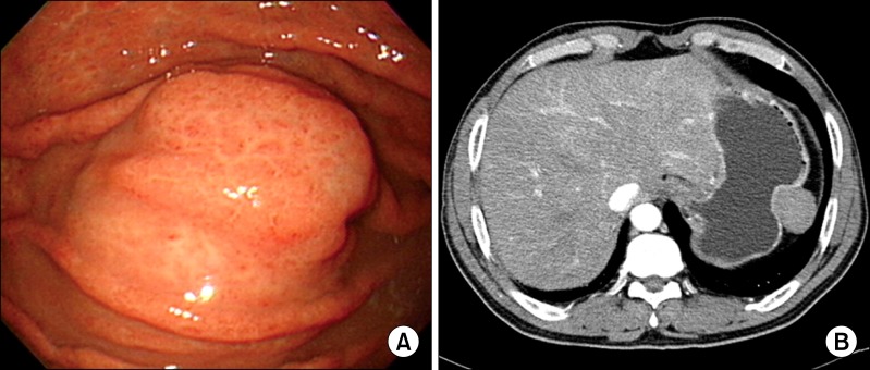
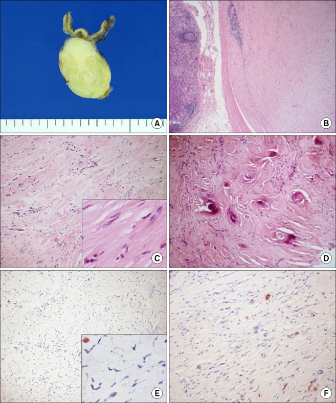
Calcifying fibrous tumor (CFT) is a rare, benign mesenchymal tumor usually affecting children and young adults, and it shows a predilection for the soft tissue and the abdominal cavity. Intrinsic visceral CFT is extremely rare and we present herein the case of a 59-year-old man with an asymptomatic gastric lesion, incidentally detected 1 month before this presentation. Thus, gastric endoscopy revealed a polypoid submucosal mass in the fundus, covered by an erythematous mucosa. The polypoid mass was a 3.9 × 2.7 cm-sized well-defined tumor located in the proper muscle, with extension to the subserosa. The tumor showed characteristic hypocellular sclerosis with coarse collagen, mononuclear inflammatory infiltrates, sparse fibroblastic spindle cells and occasional, psammomatous or dystrophic calcifications. Immunohistochemically, the spindle cells were negative for CD117, CD34, platelet-derived growth factor receptor-alpha, S100, smooth muscle actin, desmin and anaplastic lymphoma kinase.
钙化性纤维瘤(CFT)是一种罕见的良性间叶组织肿瘤,通常发生于儿童和年轻人,好发于软组织和腹腔。原发性内脏CFT极为罕见,本文报告一例59岁男性,在此次就诊前1个月偶然发现无症状的胃部病变。因此,胃镜检查发现胃底有一个息肉样黏膜下肿块,表面覆盖有红斑样黏膜。该息肉样肿块是一个大小为3.9×2.7 cm、边界清晰的肿瘤,位于固有肌层,累及浆膜下层。肿瘤表现为特征性的细胞稀少性硬化,伴有粗大胶原、单核炎性浸润、稀疏的成纤维细胞梭形细胞以及偶尔的砂粒体样或营养不良性钙化。免疫组化显示,梭形细胞CD117、CD34、血小板衍生生长因子受体α、S100、平滑肌肌动蛋白、结蛋白和间变性淋巴瘤激酶均为阴性。