• 文献检索
  • 文档翻译
  • 深度研究
  • 学术资讯
  • Suppr Zotero 插件Zotero 插件
  • 邀请有礼
  • 套餐&价格
  • 历史记录
应用&插件
Suppr Zotero 插件Zotero 插件浏览器插件Mac 客户端Windows 客户端微信小程序
定价
高级版会员购买积分包购买API积分包
服务
文献检索文档翻译深度研究API 文档MCP 服务
关于我们
关于 Suppr公司介绍联系我们用户协议隐私条款
关注我们

Suppr 超能文献

核心技术专利:CN118964589B侵权必究
粤ICP备2023148730 号-1Suppr @ 2026

文献检索

告别复杂PubMed语法,用中文像聊天一样搜索,搜遍4000万医学文献。AI智能推荐,让科研检索更轻松。

立即免费搜索

文件翻译

保留排版,准确专业,支持PDF/Word/PPT等文件格式,支持 12+语言互译。

免费翻译文档

深度研究

AI帮你快速写综述,25分钟生成高质量综述,智能提取关键信息,辅助科研写作。

立即免费体验

心外膜脂肪组织厚度是无症状 2 型糖尿病患者冠状动脉狭窄的一个指标:心脏磁共振评估。

Epicardial adipose tissue thickness is an indicator for coronary artery stenosis in asymptomatic type 2 diabetic patients: its assessment by cardiac magnetic resonance.

机构信息

Department of Internal Medicine, Yonsei University College of Medicine, Seoul, South Korea.

出版信息

Cardiovasc Diabetol. 2012 Jul 18;11:83. doi: 10.1186/1475-2840-11-83.

DOI:10.1186/1475-2840-11-83
PMID:22809408
原文链接:https://pmc.ncbi.nlm.nih.gov/articles/PMC3461492/
Abstract

BACKGROUND

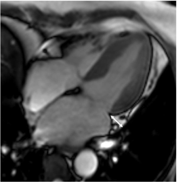
We used cardiovascular magnetic resonance (CMR) to investigate the association between epicardial adipose tissue (EAT) thickness and silent myocardial ischemia, as well as coronary artery stenosis, in asymptomatic type 2 diabetic patients.

METHODS

The study included 100 type 2 diabetic subjects (51 male and 49 female; mean age: 56 ± 7 years). Silent myocardial ischemia, as determined by CMR, was defined as evidence of inducible ischemia or myocardial infarction. Signal reduction or stenosis of ≥ 50% in the vessel diameter was used as the criteria for significant coronary artery stenosis on coronary magnetic resonance (MR) angiography.

RESULTS

EAT thickness was positively correlated with body mass index (BMI), waist-to-hip ratio, systolic blood pressure, postprandial glucose, fasting/postprandial triglyceride (TG), serum glycated hemoglobin (HbA1c) level, and homeostasis model assessment of insulin resistance (HOMA-IR) score. Significant coronary artery stenosis was found in 24 patients, while 14 patients had silent myocardial ischemia in CMR (1 with silent myocardial infarction, 11 with inducible ischemia, and 2 with both). EAT thickness was greater in patients who had coronary artery stenosis (13.0 ± 2.6 mm vs. 11.5 ± 2.1 mm, p = 0.01), but did not differ between the subjects with or without silent myocardial ischemia on CMR images (12.8 ± 2.1 vs. 11.7 ± 2.3 mm, p = 0.11). Multivariate logistic regression analysis indicated that EAT thickness was an independent indicator for significant coronary artery stenosis after adjusting for traditional risk factors (OR 1.403, p = 0.026).

CONCLUSIONS

Increased EAT thickness assessed by CMR is an independent risk factor for significant coronary artery stenosis in asymptomatic type 2 diabetes. However, EAT thickness was not associated with silent myocardial ischemia.

摘要

背景

我们使用心血管磁共振(CMR)研究了心外膜脂肪组织(EAT)厚度与无症状 2 型糖尿病患者的无症状性心肌缺血和冠状动脉狭窄之间的关系。

方法

该研究纳入了 100 例 2 型糖尿病患者(男 51 例,女 49 例;平均年龄:56±7 岁)。CMR 确定的无症状性心肌缺血定义为可诱导缺血或心肌梗死的证据。冠状动脉磁共振血管造影(CMR-Angiography)上≥50%的血管直径信号减少或狭窄被用作冠状动脉狭窄的标准。

结果

EAT 厚度与体重指数(BMI)、腰臀比、收缩压、餐后血糖、空腹/餐后甘油三酯(TG)、糖化血红蛋白(HbA1c)水平和胰岛素抵抗稳态模型评估(HOMA-IR)评分呈正相关。24 例患者存在明显的冠状动脉狭窄,14 例患者在 CMR 上有无症状性心肌缺血(1 例无症状性心肌梗死,11 例可诱导性缺血,2 例同时存在)。有冠状动脉狭窄的患者 EAT 厚度更大(13.0±2.6mm 比 11.5±2.1mm,p=0.01),但在 CMR 图像上有无无症状性心肌缺血的患者之间无差异(12.8±2.1mm 比 11.7±2.3mm,p=0.11)。多变量 logistic 回归分析表明,在校正传统危险因素后,EAT 厚度是冠状动脉显著狭窄的独立指标(OR 1.403,p=0.026)。

结论

CMR 评估的 EAT 厚度增加是无症状 2 型糖尿病患者冠状动脉显著狭窄的独立危险因素。然而,EAT 厚度与无症状性心肌缺血无关。

https://cdn.ncbi.nlm.nih.gov/pmc/blobs/e73e/3461492/ba5c094ede9b/1475-2840-11-83-1.jpg

相似文献

1
Epicardial adipose tissue thickness is an indicator for coronary artery stenosis in asymptomatic type 2 diabetic patients: its assessment by cardiac magnetic resonance.心外膜脂肪组织厚度是无症状 2 型糖尿病患者冠状动脉狭窄的一个指标:心脏磁共振评估。
Cardiovasc Diabetol. 2012 Jul 18;11:83. doi: 10.1186/1475-2840-11-83.
2
Epicardial fat volume and concurrent presence of both myocardial ischemia and obstructive coronary artery disease.心外膜脂肪体积与心肌缺血和阻塞性冠状动脉疾病同时存在。
Atherosclerosis. 2012 Apr;221(2):422-6. doi: 10.1016/j.atherosclerosis.2011.12.018. Epub 2012 Jan 4.
3
Correlation of echocardiographic epicardial fat thickness with severity of coronary artery disease in patients with acute myocardial infarction.急性心肌梗死患者超声心动图测量的心包脂肪厚度与冠状动脉疾病严重程度的相关性
Echocardiography. 2014 Nov;31(10):1177-81. doi: 10.1111/echo.12545. Epub 2014 Mar 19.
4
Epicardial adipose tissue volume and myocardial ischemia in asymptomatic people living with diabetes: a cross-sectional study.无症状糖尿病患者的心外膜脂肪组织体积与心肌缺血:一项横断面研究。
Cardiovasc Diabetol. 2021 Nov 24;20(1):224. doi: 10.1186/s12933-021-01420-5.
5
Relation between quantity and quality of peri-coronary epicardial adipose tissue and its underlying hemodynamically significant coronary stenosis.心外膜冠状脂肪组织的数量与质量及其潜在的血流动力学意义的冠状动脉狭窄之间的关系。
BMC Cardiovasc Disord. 2020 May 15;20(1):226. doi: 10.1186/s12872-020-01499-w.
6
The ratio of epicardial to body fat improves the prediction of coronary artery disease beyond calcium and Framingham risk scores.心外膜脂肪与体脂的比例,相比钙化积分和弗雷明汉风险评分,能更好地预测冠状动脉疾病。
Int J Cardiovasc Imaging. 2016 Jun;32 Suppl 1:117-27. doi: 10.1007/s10554-016-0912-2. Epub 2016 Jun 13.
7
N-terminal fragment of pro-brain natriuretic peptide (NT-proBNP) for predicting silent myocardial ischaemia in type 2 diabetes mellitus independent of microalbuminuria.脑钠肽前体 N 端片段(NT-proBNP)预测 2 型糖尿病患者无症状性心肌缺血,与微量白蛋白尿无关。
Diabetes Metab Res Rev. 2010 Oct;26(7):534-9. doi: 10.1002/dmrr.1113.
8
Assessing the Relation of Epicardial Fat Thickness and Volume, Quantified by 256-Slice Computed Tomography Scan, With Coronary Artery Disease and Cardiovascular Risk Factors.通过256层计算机断层扫描评估心外膜脂肪厚度和体积与冠状动脉疾病及心血管危险因素之间的关系。
Arch Iran Med. 2018 Mar 1;21(3):95-100.
9
Epicardial adipose tissue as a predictor of coronary artery disease in asymptomatic subjects.心外膜脂肪组织作为无症状患者冠状动脉疾病的预测因子。
Am J Cardiol. 2012 Aug 15;110(4):534-8. doi: 10.1016/j.amjcard.2012.04.024. Epub 2012 May 10.
10
The relation of location-specific epicardial adipose tissue thickness and obstructive coronary artery disease: systemic review and meta-analysis of observational studies.特定部位的心外膜脂肪组织厚度与阻塞性冠状动脉疾病的关系:观察性研究的系统评价和荟萃分析
BMC Cardiovasc Disord. 2014 May 4;14:62. doi: 10.1186/1471-2261-14-62.

引用本文的文献

1
The Role of Epicardial Adipose Tissue in Acute Coronary Syndromes, Post-Infarct Remodeling and Cardiac Regeneration.心外膜脂肪组织在急性冠状动脉综合征、心肌梗死后重构和心脏再生中的作用。
Int J Mol Sci. 2024 Mar 22;25(7):3583. doi: 10.3390/ijms25073583.
2
Role of epicardial adipose tissue in diabetic cardiomyopathy through the lens of cardiovascular magnetic resonance imaging - a narrative review.从心血管磁共振成像角度看心外膜脂肪组织在糖尿病性心肌病中的作用——一篇叙述性综述
Ther Adv Endocrinol Metab. 2024 Mar 10;15:20420188241229540. doi: 10.1177/20420188241229540. eCollection 2024.
3
The Role of Epicardial Adipose Tissue in the Development of Atrial Fibrillation, Coronary Artery Disease and Chronic Heart Failure in the Context of Obesity and Type 2 Diabetes Mellitus: A Narrative Review.

本文引用的文献

1
Cardiovascular Risk Assessment with Vascular Function, Carotid Atherosclerosis and the UKPDS Risk Engine in Korean Patients with Newly Diagnosed Type 2 Diabetes.韩国新诊断 2 型糖尿病患者的血管功能、颈动脉粥样硬化和 UKPDS 风险引擎的心血管风险评估。
Diabetes Metab J. 2011 Dec;35(6):619-27. doi: 10.4093/dmj.2011.35.6.619. Epub 2011 Dec 26.
2
Association between EPCs count and rate of coronary revascularization in asymptomatic type 2 diabetic patients.在无症状 2 型糖尿病患者中,EPCs 计数与冠状动脉血运重建率的相关性。
Acta Diabetol. 2012 Dec;49(6):413-20. doi: 10.1007/s00592-011-0360-3. Epub 2011 Dec 13.
3
Association of chemerin mRNA expression in human epicardial adipose tissue with coronary atherosclerosis.
肥胖与2型糖尿病背景下,心外膜脂肪组织在房颤、冠状动脉疾病和慢性心力衰竭发生发展中的作用:一项叙述性综述
J Cardiovasc Dev Dis. 2022 Jul 5;9(7):217. doi: 10.3390/jcdd9070217.
4
Incremental Prognostic Value of Pericoronary Adipose Tissue Thickness Measured Using Cardiac Magnetic Resonance Imaging After Revascularization in Patients With ST-Elevation Myocardial Infarction.采用心脏磁共振成像测量的ST段抬高型心肌梗死患者血运重建后冠状动脉周围脂肪组织厚度的增量预后价值
Front Cardiovasc Med. 2022 Mar 4;9:781402. doi: 10.3389/fcvm.2022.781402. eCollection 2022.
5
Epicardial adipose tissue volume and myocardial ischemia in asymptomatic people living with diabetes: a cross-sectional study.无症状糖尿病患者的心外膜脂肪组织体积与心肌缺血:一项横断面研究。
Cardiovasc Diabetol. 2021 Nov 24;20(1):224. doi: 10.1186/s12933-021-01420-5.
6
Epicardial adipose tissue: an emerging biomarker of cardiovascular complications in type 2 diabetes?心外膜脂肪组织:2型糖尿病心血管并发症的一种新兴生物标志物?
Ther Adv Endocrinol Metab. 2020 May 25;11:2042018820928824. doi: 10.1177/2042018820928824. eCollection 2020.
7
Clinical Use of Cardiac Magnetic Resonance in Systemic Heart Disease.心脏磁共振成像在全身性心脏病中的临床应用
Eur Cardiol. 2014 Jul;9(1):21-27. doi: 10.15420/ecr.2014.9.1.21.
8
Is echocardiographic epicardial fat thickness increased in patients with coronary artery disease? A systematic review and meta-analysis.冠心病患者的超声心动图心包脂肪厚度会增加吗?一项系统评价和荟萃分析。
Electron Physician. 2018 Sep 9;10(9):7249-7258. doi: 10.19082/7249. eCollection 2018 Sep.
9
Clinical importance of epicardial adipose tissue.心外膜脂肪组织的临床重要性。
Arch Med Sci. 2017 Jun;13(4):864-874. doi: 10.5114/aoms.2016.63259. Epub 2016 Oct 26.
10
Investigating interactions between epicardial adipose tissue and cardiac myocytes: what can we learn from different approaches?探讨心外膜脂肪组织与心肌细胞之间的相互作用:不同方法能为我们带来哪些启示?
Br J Pharmacol. 2017 Oct;174(20):3542-3560. doi: 10.1111/bph.13678. Epub 2017 Jan 22.
人心外膜脂肪组织中 chemerin mRNA 表达与冠状动脉粥样硬化的关系。
Cardiovasc Diabetol. 2011 Oct 7;10:87. doi: 10.1186/1475-2840-10-87.
4
Postprandial Triglyceride Is Associated with Fasting Triglyceride and HOMA-IR in Korean Subjects with Type 2 Diabetes.韩国 2 型糖尿病患者餐后甘油三酯与空腹甘油三酯和 HOMA-IR 相关。
Diabetes Metab J. 2011 Aug;35(4):404-10. doi: 10.4093/dmj.2011.35.4.404. Epub 2011 Aug 31.
5
Increased epicardial fat tissue is a marker of metabolic syndrome in adult patients.心外膜脂肪组织增加是成年患者代谢综合征的一个标志物。
Int J Cardiol. 2013 May 10;165(2):308-13. doi: 10.1016/j.ijcard.2011.08.067. Epub 2011 Sep 16.
6
Cardiac 64-multislice computed tomography reveals increased epicardial fat volume in patients with acute coronary syndrome.心脏 64 层计算机断层扫描显示急性冠状动脉综合征患者的心外膜脂肪体积增加。
Am J Cardiol. 2011 Oct 15;108(8):1119-23. doi: 10.1016/j.amjcard.2011.06.012. Epub 2011 Aug 1.
7
Pericardial rather than epicardial fat is a cardiometabolic risk marker: an MRI vs echo study.心包脂肪而非心外膜脂肪是心血管代谢风险标志物:一项 MRI 与超声研究。
J Am Soc Echocardiogr. 2011 Oct;24(10):1156-62. doi: 10.1016/j.echo.2011.06.013. Epub 2011 Jul 26.
8
MRI measured epicardial adipose tissue thickness at the right AV groove differentiates inflammatory status in obese men with metabolic syndrome.MRI 测量右房室沟处的心外膜脂肪组织厚度可区分合并代谢综合征的肥胖男性的炎症状态。
Obesity (Silver Spring). 2012 Mar;20(3):525-32. doi: 10.1038/oby.2011.155. Epub 2011 Jun 9.
9
Feasibility of dual gradient-echo in-phase and opposed-phase magnetic resonance imaging for the evaluation of epicardial fat.双梯度回波同相位和反相位磁共振成像评估心外膜脂肪的可行性
Acta Radiol. 2011 Sep 1;52(7):723-9. doi: 10.1258/ar.2011.100454. Epub 2011 May 17.
10
Epicardial adipose tissue: an emerging role for the development of coronary atherosclerosis.心外膜脂肪组织:在冠状动脉粥样硬化发展中的新作用。
Clin Cardiol. 2011 Mar;34(3):143-4. doi: 10.1002/clc.20893.