Division of Biology and Medicine, Department of Molecular Microbiology and Immunology and Warren Alpert Medical School, Brown University, Providence, Rhode Island, United States of America.
PLoS One. 2012;7(8):e42850. doi: 10.1371/journal.pone.0042850. Epub 2012 Aug 3.
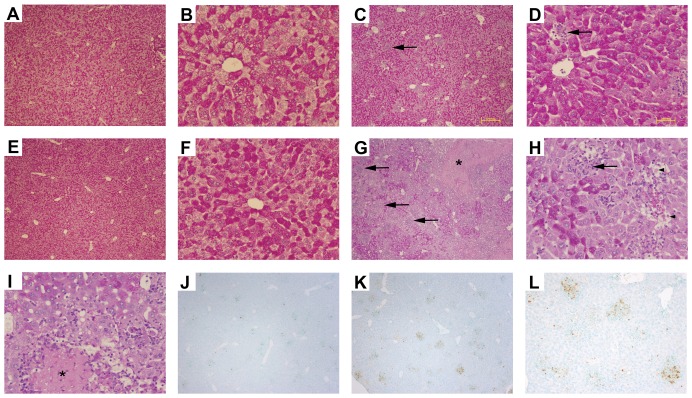
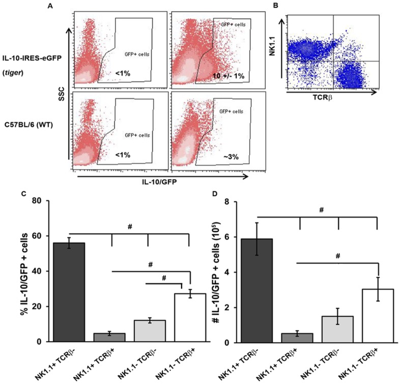
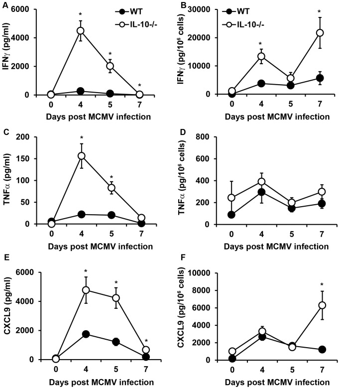
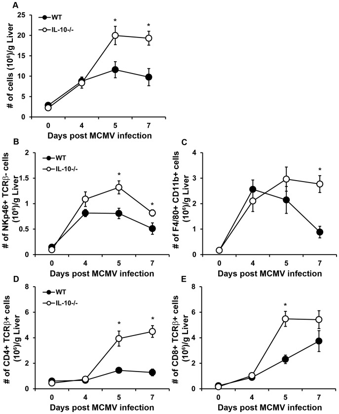
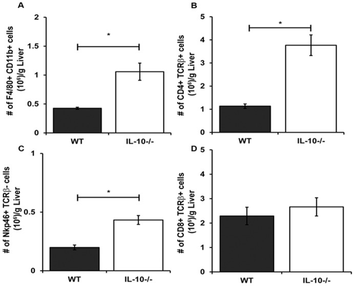
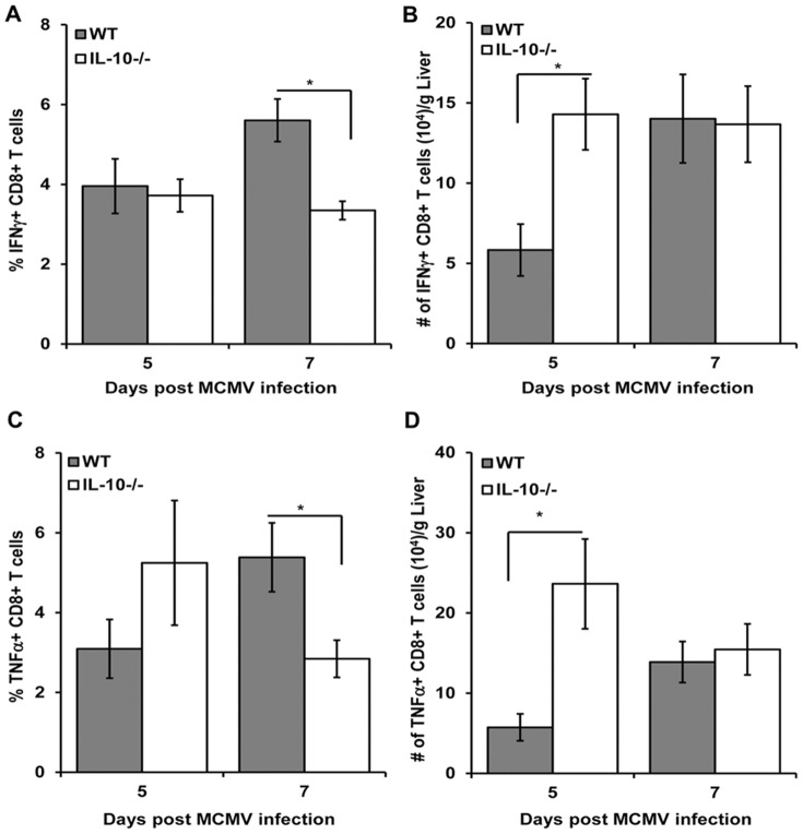
Various cell types in both lymphoid and non-lymphoid tissues produce the anti-inflammatory cytokine interleukin (IL)-10 during murine cytomegalovirus (MCMV) infection. The functions of IL-10 in the liver during acute infection and the cells that generate this cytokine at this site have not been extensively investigated. In this study, we demonstrate that the production of IL-10 in the liver is elevated in C57BL/6 mice during late acute MCMV infection. Using IL-10 green fluorescence protein (GFP) reporter knock-in mice, designated IL-10-internal ribosomal entry site (IRES)-GFP-enhanced reporter (tiger), NK cells are identified as major IL-10 expressing cells in the liver after infection, along with T cells and other leukocytes. In the absence of IL-10, mice exhibit marked elevations in proinflammatory cytokines and in the numbers of mononuclear cells and lymphocytes infiltrating the liver during this infection. IL-10-deficiency also enhances liver injury without improving viral clearance from this site. Collectively, the results indicate that IL-10-producing cells in the liver provide protection from collateral injury by modulating the inflammatory response associated with MCMV infection.
在小鼠巨细胞病毒(MCMV)感染期间,淋巴组织和非淋巴组织中的各种细胞类型都会产生抗炎细胞因子白细胞介素(IL)-10。在急性感染期间,IL-10 在肝脏中的功能以及在该部位产生这种细胞因子的细胞尚未得到广泛研究。在这项研究中,我们证明在 C57BL/6 小鼠的急性 MCMV 感染后期,肝脏中 IL-10 的产生增加。使用 IL-10 绿色荧光蛋白(GFP)报告基因敲入小鼠,命名为 IL-10 内部核糖体进入位点(IRES)-GFP 增强报告基因(tiger),发现 NK 细胞是感染后肝脏中主要的 IL-10 表达细胞,与 T 细胞和其他白细胞一起。在缺乏 IL-10 的情况下,与感染期间单核细胞和淋巴细胞浸润肝脏相关的促炎细胞因子和单核细胞数量显著增加。IL-10 缺乏也会增强肝损伤,而不会改善该部位的病毒清除。总之,这些结果表明,肝脏中的 IL-10 产生细胞通过调节与 MCMV 感染相关的炎症反应提供了对继发损伤的保护。