Neuroimmunology Laboratory, Center for Infectious Diseases and Microbiology Translational Research, Department of Medicine, University of Minnesota, Minneapolis, MN 55455, USA.
J Neurovirol. 2011 Oct;17(5):424-37. doi: 10.1007/s13365-011-0042-5. Epub 2011 Jul 29.
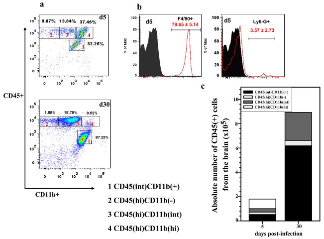
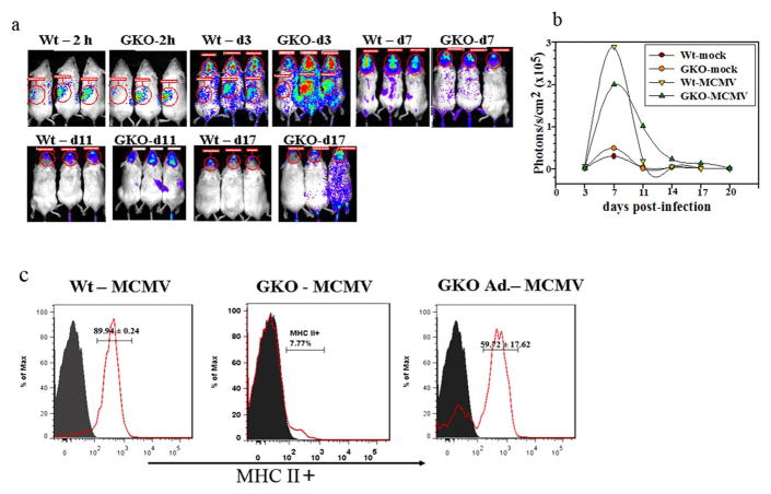
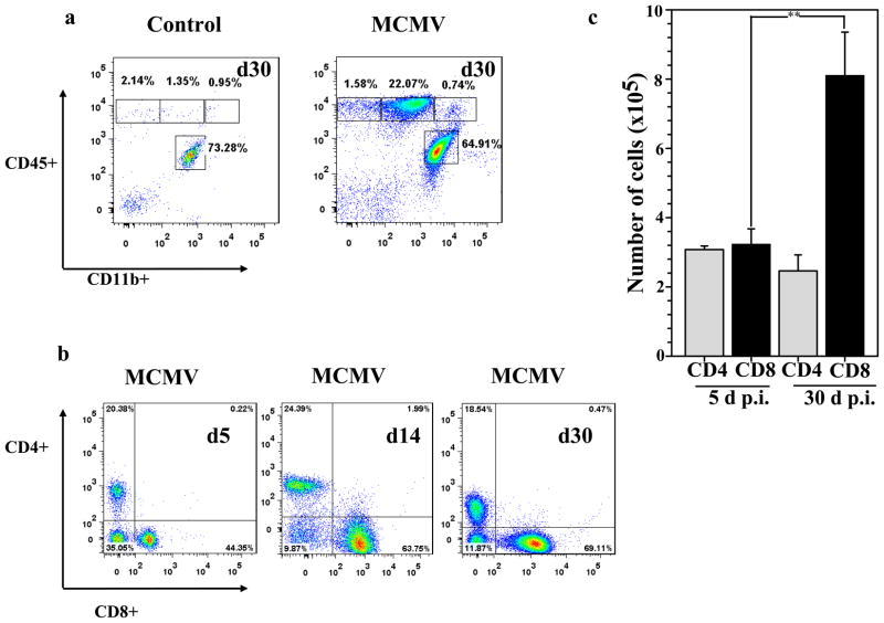
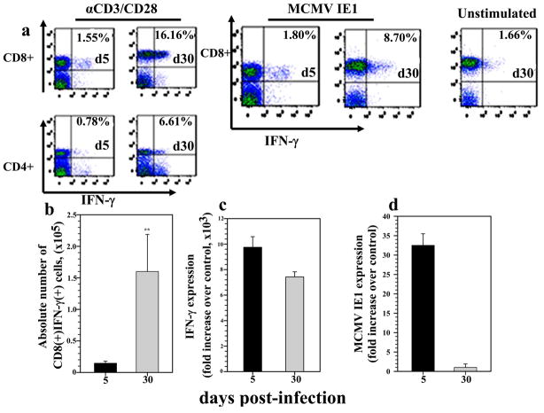
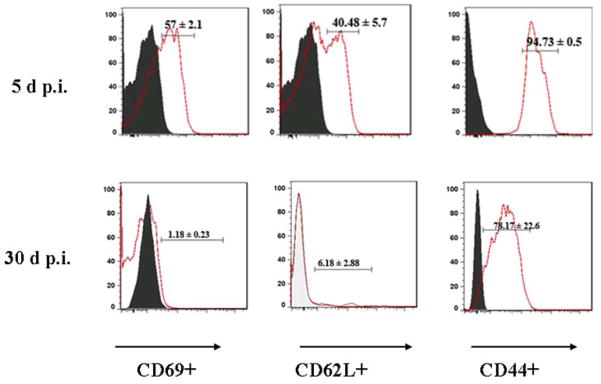
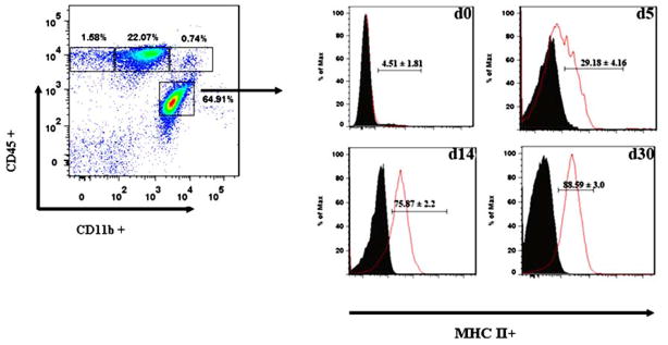
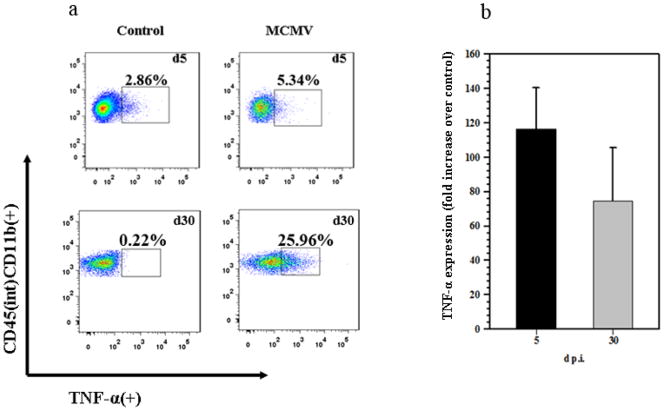
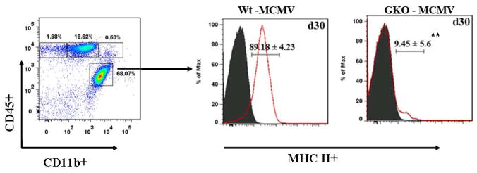
Murine cytomegalovirus (MCMV) brain infection stimulates microglial cell-driven proinflammatory chemokine production which precedes the presence of brain-infiltrating systemic immune cells. Here, we show that in response to MCMV brain infection, antigen-specific CD8(+) T cells migrated into the brain and persisted as long-lived memory cells. The role of these persistent T cells in the brain is unclear because most of our understanding of antimicrobial T cell responses comes from analyses of lymphoid tissue. Strikingly, memory T cells isolated from the brain exhibited an effector phenotype and produced IFN-γ upon restimulation with viral peptide. Furthermore, we observed time-dependent and long-term activation of resident microglia, indicated by chronic MHC class II up-regulation and TNF-α production. The immune response in this immunologically restricted site persisted in the absence of active viral replication. Lymphocyte infiltrates were detected until 30 days post-infection (p.i.), with CD8(+) and CD4(+) T cells present at a 3:1 ratio, respectively. We then investigated the role of IFN-γ in chronic microglial activation by using IFN-γ-knockout (GKO) mice. At 30 days p.i., GKO mice demonstrated a similar phenotypic brain infiltrate when compared to wild-type mice (Wt), however, MHC class II expression on microglia isolated from these GKO mice was significantly lower compared to Wt animals. When IFN-γ producing CD8(+) T cells were reconstituted in GKO mice, MHC class II up-regulation on microglial cells was restored. Taken together, these results suggest that MCMV brain infection results in long-term persistence of antigen-specific CD8(+) T cells which produce IFN-γ and drive chronic microglial cell activation. This response was found to be dependent on IFN-γ production by viral Ag-specific T cells during the chronic phase of disease.
鼠巨细胞病毒(MCMV)脑感染刺激小胶质细胞驱动的促炎趋化因子产生,这先于脑浸润的系统性免疫细胞的出现。在这里,我们表明,针对 MCMV 脑感染,抗原特异性 CD8(+) T 细胞迁移到大脑中,并作为长期记忆细胞持续存在。这些持久的 T 细胞在大脑中的作用尚不清楚,因为我们对抗菌 T 细胞反应的大部分理解来自对淋巴组织的分析。引人注目的是,从大脑中分离出的记忆 T 细胞表现出效应表型,并在受到病毒肽刺激时产生 IFN-γ。此外,我们观察到常驻小胶质细胞的时间依赖性和长期激活,表现为 MHC 类 II 持续上调和 TNF-α 产生。在没有活跃病毒复制的情况下,免疫反应在这个免疫受限的部位持续存在。淋巴细胞浸润可检测到感染后 30 天(p.i.),CD8(+) 和 CD4(+) T 细胞的比例分别为 3:1。然后,我们通过使用 IFN-γ 敲除(GKO)小鼠研究 IFN-γ 在慢性小胶质细胞激活中的作用。在 30 天 p.i.时,与野生型(Wt)小鼠相比,GKO 小鼠表现出相似的表型脑浸润,但从这些 GKO 小鼠分离的小胶质细胞上的 MHC 类 II 表达明显低于 Wt 动物。当 IFN-γ 产生的 CD8(+) T 细胞在 GKO 小鼠中重建时,小胶质细胞上的 MHC 类 II 上调得到恢复。总之,这些结果表明,MCMV 脑感染导致抗原特异性 CD8(+) T 细胞的长期持续存在,这些细胞产生 IFN-γ 并驱动慢性小胶质细胞激活。发现这种反应依赖于疾病慢性期病毒 Ag 特异性 T 细胞的 IFN-γ 产生。