Department of Anatomy and Physiology, Faculty of Veterinary Science, University of Pretoria, Private Bag X04, Onderstepoort, 0110, South Africa.
Front Zool. 2012 Aug 21;9(1):21. doi: 10.1186/1742-9994-9-21.
Tonsils are secondary lymphoid organs located in the naso- and oropharynx of most mammalian species. Most tonsils are characterised by crypts surrounded by dense lymphoid tissue. However, tonsils without crypts have also been recognised. Gut-associated lymphoid tissue (GALT), although not well-organised and lacking tonsillar crypts, is abundant in the avian oropharynx and has been referred to as the "pharyngeal tonsil". In this context the pharyngeal folds present in the oropharynx of ratites have erroneously been named the pharyngeal tonsils. This study distinguishes between the different types and arrangements of lymphoid tissue in the pharyngeal region of D. novaehollandiae and S. camelus and demonstrates that both species possess a true pharyngeal tonsil which fits the classical definition of tonsils in mammals.
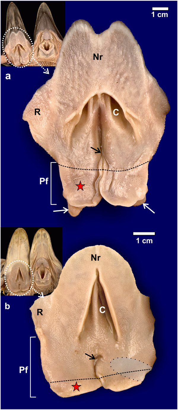
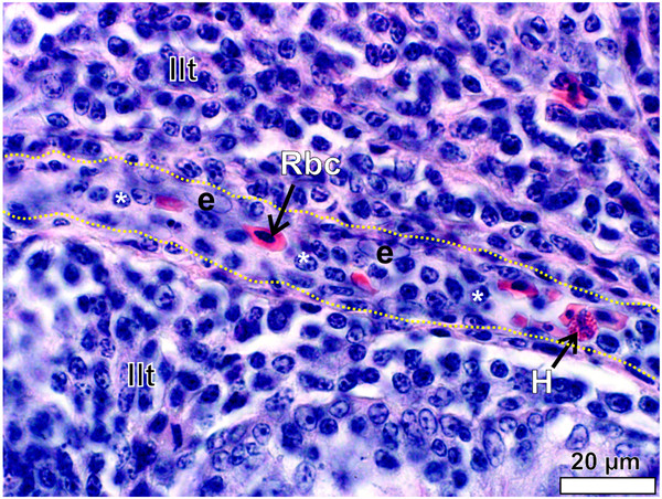
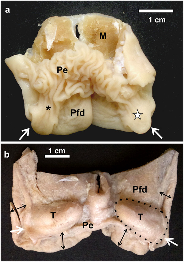
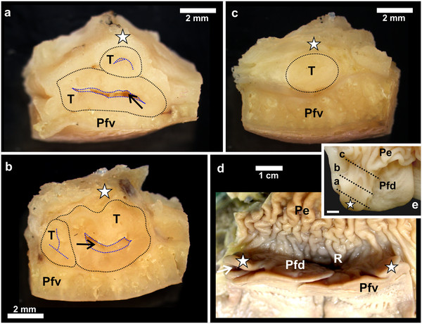
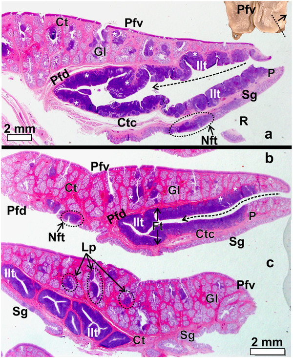
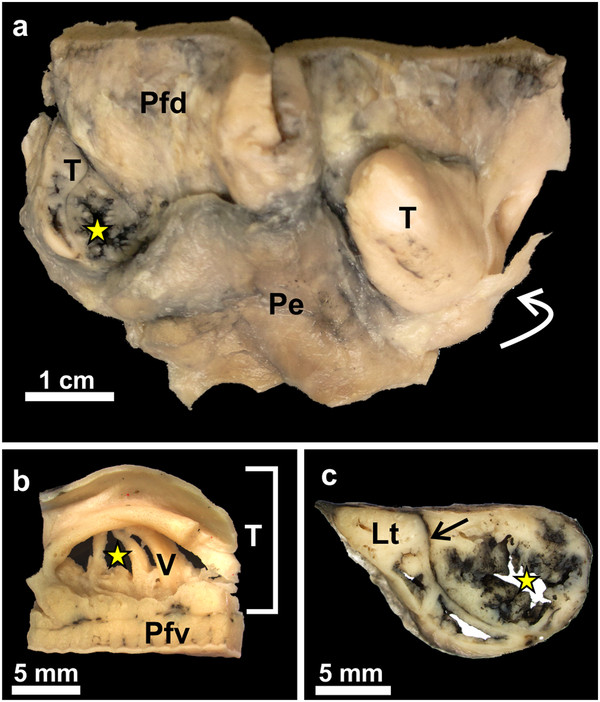
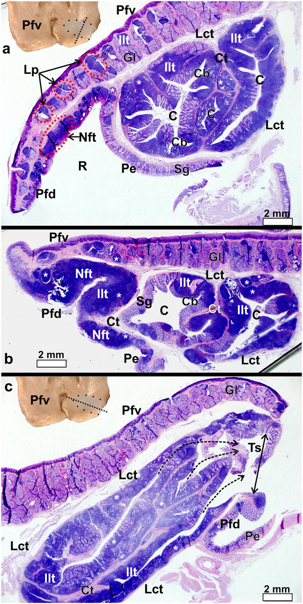
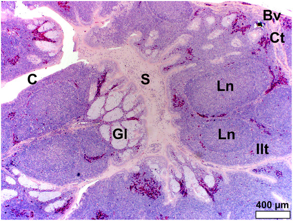
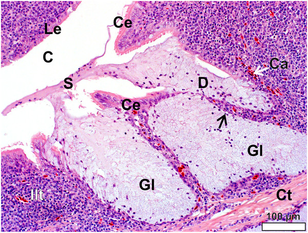
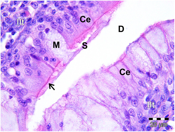
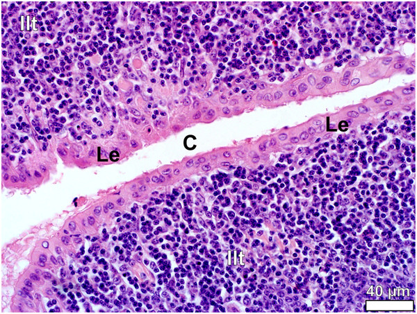
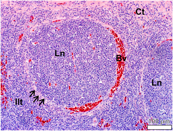
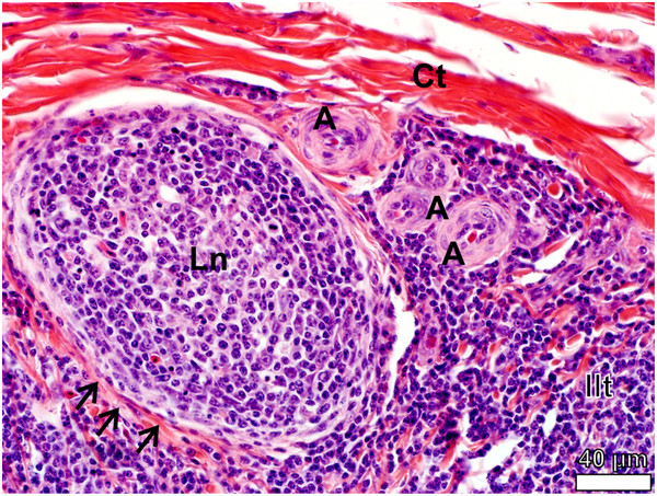
The pharyngeal tonsil (Tonsilla pharyngea) of D. novaehollandiae was located on the dorsal free surface of the pharyngeal folds and covered by a small caudo-lateral extension of the folds whereas in S. camelus the tonsil was similarly located on the dorsal surface of the pharyngeal folds but was positioned retropharyngeally and encapsulated by loose connective tissue. The pharyngeal tonsil in both species was composed of lymph nodules, inter-nodular lymphoid tissue, mucus glands, crypts and intervening connective tissue septa. In S. camelus a shallow tonsillar sinus was present. Aggregated lymph nodules and inter-nodular lymphoid tissue was associated with the mucus glands on the ventral surface of the pharyngeal folds in both species and represented the Lymphonoduli pharyngeales. Similar lymphoid tissue, but more densely packed and situated directly below the epithelium, was present on the dorsal, free surface of the pharyngeal folds and represented a small, non-follicular tonsil.
The follicular pharyngeal tonsils in D. novaehollandiae and S. camelus are distinct from the pharyngeal folds in these species and perfectly fit the classical mammalian definition of pharyngeal tonsils. The presence of a true pharyngeal tonsil differentiates these two ratite species from other known avian species where similar structures have not been described. The pharyngeal tonsils in these ratites may pose a suitable and easily accessible site for immune response surveillance as indicated by swelling and inflammation of the tonsillar tissue and pharyngeal folds. This would be facilitated by the fact that the heads of these commercially slaughtered ratites are discarded, thus sampling at these sites would not result in financial losses.
扁桃体是位于大多数哺乳动物的鼻腔和口咽的次级淋巴器官。大多数扁桃体的特征是被致密的淋巴组织包围的隐窝。然而,也有被识别出没有隐窝的扁桃体。肠道相关淋巴组织(GALT),虽然组织不发达且缺乏扁桃体隐窝,但在禽类的口咽中非常丰富,并被称为“咽扁桃体”。在这种情况下,在平胸鸟类的口咽中出现的咽皱襞错误地被命名为咽扁桃体。本研究区分了 D. novaehollandiae 和 S. camelus 咽区的不同类型和排列的淋巴组织,并证明这两个物种都具有真正的咽扁桃体,符合哺乳动物扁桃体的经典定义。
D. novaehollandiae 的咽扁桃体(Tonsilla pharyngea)位于咽皱襞的背侧游离表面,并被皱襞的小尾侧向延伸所覆盖,而在 S. camelus 中,扁桃体同样位于咽皱襞的背侧表面,但位于咽后,并被疏松的结缔组织包裹。这两个物种的咽扁桃体由淋巴小结、小结间淋巴组织、粘液腺、隐窝和中间的结缔组织隔组成。在 S. camelus 中存在一个浅的扁桃体窦。在两个物种的咽皱襞腹侧表面,聚集的淋巴小结和小结间淋巴组织与粘液腺相关,代表咽淋巴小结(Lymphonoduli pharyngeales)。在咽皱襞的背侧游离表面也存在类似的淋巴组织,但更密集地排列并直接位于上皮下方,代表一个小的、无滤泡的扁桃体。
D. novaehollandiae 和 S. camelus 的滤泡性咽扁桃体与这些物种的咽皱襞明显不同,完全符合经典的哺乳动物咽扁桃体的定义。真正的咽扁桃体的存在使这两个平胸鸟类物种与其他已知的禽类物种区分开来,后者没有描述类似的结构。这些平胸鸟类的咽扁桃体可能是一个合适且易于接近的免疫反应监测部位,因为扁桃体组织和咽皱襞的肿胀和炎症表明了这一点。事实上,这些商业屠宰的平胸鸟类的头部被丢弃,因此在这些部位采样不会导致经济损失,这使得这种情况更加便利。