Izatt Maree T, Adam Clayton J, Verzin Eugene J, Labrom Robert D, Askin Geoffrey N
Paediatric Spine Research Group, Queensland University of Technology and Mater Health Services Brisbane Ltd, Queensland, Australia.
Scoliosis. 2012 Aug 22;7(1):15. doi: 10.1186/1748-7161-7-15.
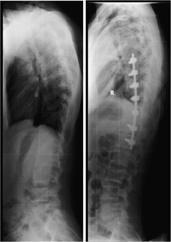
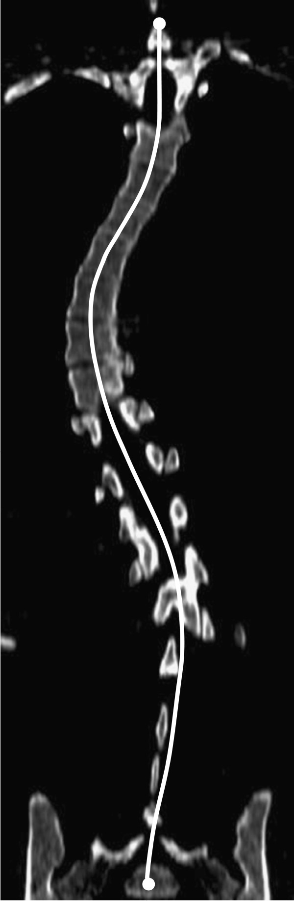
Previous studies report an increase in thoracic kyphosis after anterior approaches and a flattening of sagittal contours following posterior approaches. Difficulties with measuring sagittal parameters on radiographs are avoided with reformatted sagittal CT reconstructions due to the superior endplate clarity afforded by this imaging modality.
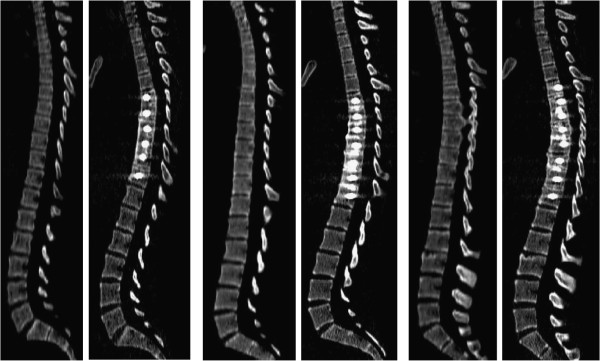
A prospective study of 30 Lenke 1 adolescent idiopathic scoliosis (AIS) patients receiving selective thoracoscopic anterior spinal fusion (TASF) was performed. Participants had ethically approved low dose CT scans at minimum 24 months after surgery in addition to their standard care following surgery. The change in sagittal contours on supine CT was compared to standing radiographic measurements of the same patients and with previous studies. Inter-observer variability was assessed as well as whether hypokyphotic and normokyphotic patient groups responded differently to the thoracoscopic anterior approach.
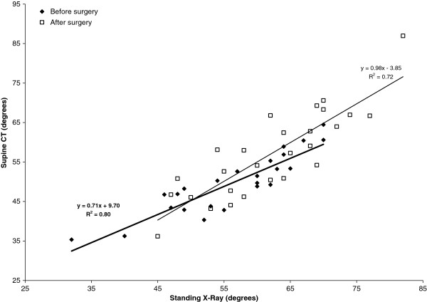
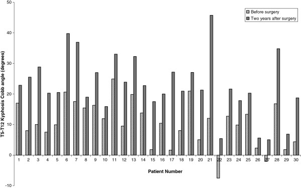
Mean T5-12 kyphosis Cobb angle increased by 11.8 degrees and lumbar lordosis increased by 5.9 degrees on standing radiographs two years after surgery. By comparison, CT measurements of kyphosis and lordosis increased by 12.3 degrees and 7.0 degrees respectively. 95% confidence intervals for inter-observer variability of sagittal contour measurements on supine CT ranged between 5-8 degrees. TASF had a slightly greater corrective effect on patients who were hypokyphotic before surgery compared with those who were normokyphotic.
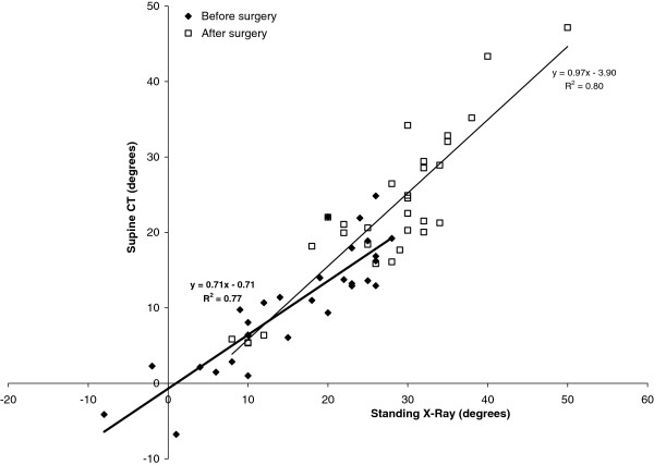
Restoration of sagittal profile is an important goal of scoliosis surgery, but reliable measurement with radiographs suffers from poor endplate clarity. TASF significantly improves thoracic kyphosis and lumbar lordosis while preserving proximal and distal junctional alignment in thoracic AIS patients. Supine CT allows greater endplate clarity for sagittal Cobb measurements and linear relationships were found between supine CT and standing radiographic measurements. In this study, improvements in sagittal kyphosis and lordosis following surgery were in agreement with prior anterior surgery studies, and add to the current evidence suggesting that anterior correction is more capable than posterior approaches of addressing the sagittal component of both the instrumented and adjacent non instrumented segments following surgical correction of progressive Lenke 1 idiopathic scoliosis.
既往研究报道,前路手术后胸椎后凸增加,后路手术后矢状面轮廓变平。由于矢状面CT重建能提供更清晰的终板图像,避免了在X线片上测量矢状面参数的困难。
对30例接受选择性胸腔镜前路脊柱融合术(TASF)的Lenke 1型青少年特发性脊柱侧凸(AIS)患者进行了一项前瞻性研究。参与者在术后至少24个月时,除了接受标准护理外,还接受了符合伦理规范的低剂量CT扫描。将仰卧位CT上矢状面轮廓的变化与同一患者的站立位X线测量结果以及既往研究进行比较。评估了观察者间的变异性,以及后凸不足和正常后凸患者组对胸腔镜前路手术的反应是否不同。
术后两年,站立位X线片上T5-12节段的平均后凸Cobb角增加了11.8度,腰椎前凸增加了5.9度。相比之下,CT测量的后凸和前凸分别增加了12.3度和7.0度。仰卧位CT上矢状面轮廓测量的观察者间变异性的95%置信区间在5-8度之间。与正常后凸患者相比,TASF对术前存在后凸不足的患者具有略大的矫正效果。
矢状面轮廓的恢复是脊柱侧凸手术的一个重要目标,但X线片测量的可靠性受终板清晰度差的影响。TASF显著改善了胸椎后凸和腰椎前凸,同时保持了胸椎AIS患者近端和远端交界区的对线。仰卧位CT能更清晰地显示终板,用于矢状面Cobb角测量,并且发现仰卧位CT与站立位X线测量之间存在线性关系。在本研究中,术后矢状面后凸和前凸的改善与既往前路手术研究结果一致,并且补充了当前的证据,表明在Lenke 1型进展性特发性脊柱侧凸手术矫正后,前路矫正比后路手术更能解决融合节段及相邻非融合节段的矢状面问题。