• 文献检索
  • 文档翻译
  • 深度研究
  • 学术资讯
  • Suppr Zotero 插件Zotero 插件
  • 邀请有礼
  • 套餐&价格
  • 历史记录
应用&插件
Suppr Zotero 插件Zotero 插件浏览器插件Mac 客户端Windows 客户端微信小程序
定价
高级版会员购买积分包购买API积分包
服务
文献检索文档翻译深度研究API 文档MCP 服务
关于我们
关于 Suppr公司介绍联系我们用户协议隐私条款
关注我们

Suppr 超能文献

核心技术专利:CN118964589B侵权必究
粤ICP备2023148730 号-1Suppr @ 2026

文献检索

告别复杂PubMed语法,用中文像聊天一样搜索,搜遍4000万医学文献。AI智能推荐,让科研检索更轻松。

立即免费搜索

文件翻译

保留排版,准确专业,支持PDF/Word/PPT等文件格式,支持 12+语言互译。

免费翻译文档

深度研究

AI帮你快速写综述,25分钟生成高质量综述,智能提取关键信息,辅助科研写作。

立即免费体验

缝隙连接晶格图像的相关性分析。

Correlation analysis of gap junction lattice images.

作者信息

Sosinsky G E, Baker T S, Caspar D L, Goodenough D A

机构信息

Rosenstiel Basic Medical Sciences Research Center, Brandeis University, Waltham, Massachusetts 02254-9110.

出版信息

Biophys J. 1990 Nov;58(5):1213-26. doi: 10.1016/S0006-3495(90)82462-0.

DOI:10.1016/S0006-3495(90)82462-0
PMID:2291941
原文链接:https://pmc.ncbi.nlm.nih.gov/articles/PMC1281066/
Abstract

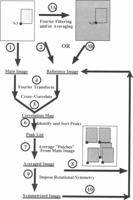
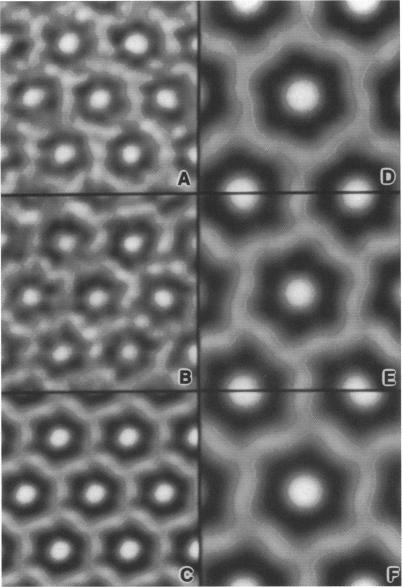
Fourier averages of connexon images computed from low-irradiation electron micrographs of isolated negatively stained gap junction domains exhibited differences in stain distribution and connexon orientation. To analyze these polymorphic structures, correlation averaging methods were applied to images from negatively stained and frozen-hydrated specimens. For the negatively stained specimens, separate averages over two subsets of connexons with differing degrees of stain accumulation in the axial channel were obtained. Two populations of connexons with opposite skew orientations were distinguishable within a single junctional domain of a frozen-hydrated specimen. Correlation maps calculated using the left- and right-skewed references showed that the selected connexons tend to locally cluster. Using correlation methods to analyze packing disorder in a typical connexon lattice, we estimated the root-mean-square variation in the nearest neighbor pair separation to be approximately 11% of the lattice constant. Displacements of the connexons relative to each other increased with increasing pair separation in the lattice, rather like a liquid, although long-range orientation order was conserved as in a crystal. These results support the hypothesis that the hexagonal ordering of the connexons results from short-range repulsive forces.

摘要

从分离的经负染色的间隙连接域的低辐射电子显微照片计算得到的连接子图像的傅里叶平均值,在染色分布和连接子取向上表现出差异。为了分析这些多态结构,相关平均方法被应用于来自负染色和冷冻水合标本的图像。对于负染色标本,在轴向通道中具有不同程度染色积累的连接子的两个子集上分别进行了平均。在冷冻水合标本的单个连接域内,可以区分出具有相反倾斜取向的两类连接子。使用左斜和右斜参考计算的相关图表明,所选连接子倾向于局部聚集。通过相关方法分析典型连接子晶格中的堆积无序,我们估计最近邻对间距的均方根变化约为晶格常数的11%。连接子相对于彼此的位移随着晶格中对间距的增加而增加,有点像液体,尽管像晶体一样保持着长程取向有序。这些结果支持了连接子的六边形排列是由短程排斥力导致的这一假设。

https://cdn.ncbi.nlm.nih.gov/pmc/blobs/c471/1281066/ec706dadcad9/biophysj00121-0143-a.jpg
https://cdn.ncbi.nlm.nih.gov/pmc/blobs/c471/1281066/5f7cd6864b8c/biophysj00121-0134-a.jpg
https://cdn.ncbi.nlm.nih.gov/pmc/blobs/c471/1281066/0aaef3a07ac0/biophysj00121-0136-a.jpg
https://cdn.ncbi.nlm.nih.gov/pmc/blobs/c471/1281066/5e13dce964cc/biophysj00121-0137-a.jpg
https://cdn.ncbi.nlm.nih.gov/pmc/blobs/c471/1281066/d6fdc781cf88/biophysj00121-0139-a.jpg
https://cdn.ncbi.nlm.nih.gov/pmc/blobs/c471/1281066/2ddc09918e3c/biophysj00121-0140-a.jpg
https://cdn.ncbi.nlm.nih.gov/pmc/blobs/c471/1281066/fb2c9f63a524/biophysj00121-0141-a.jpg
https://cdn.ncbi.nlm.nih.gov/pmc/blobs/c471/1281066/9ed4f12cf19e/biophysj00121-0142-a.jpg
https://cdn.ncbi.nlm.nih.gov/pmc/blobs/c471/1281066/ec706dadcad9/biophysj00121-0143-a.jpg

相似文献

1
Correlation analysis of gap junction lattice images.缝隙连接晶格图像的相关性分析。
Biophys J. 1990 Nov;58(5):1213-26. doi: 10.1016/S0006-3495(90)82462-0.
2
Gap junction structures. IV. Asymmetric features revealed by low-irradiation microscopy.间隙连接结构。IV. 低辐射显微镜揭示的不对称特征。
J Cell Biol. 1983 Jan;96(1):204-16. doi: 10.1083/jcb.96.1.204.
3
Gap junction structures. VII. Analysis of connexon images obtained with cationic and anionic negative stains.间隙连接结构。VII. 用阳离子和阴离子负染获得的连接子图像分析。
J Mol Biol. 1985 Jul 5;184(1):81-98. doi: 10.1016/0022-2836(85)90045-2.
4
Organization of connexons in isolated rat liver gap junctions.分离的大鼠肝脏间隙连接中连接子的组织形式
Biophys J. 1988 Jul;54(1):105-12. doi: 10.1016/S0006-3495(88)82935-7.
5
Gap junction structures. VIII. Membrane cross-sections.间隙连接结构。VIII. 膜横截面。
Biophys J. 1988 May;53(5):709-22. doi: 10.1016/S0006-3495(88)83152-7.
6
Image analysis of gap junction structures.
Electron Microsc Rev. 1992;5(1):59-76. doi: 10.1016/0892-0354(92)90005-b.
7
Structure of the extracellular surface of the gap junction by atomic force microscopy.通过原子力显微镜观察间隙连接细胞外表面的结构
Biophys J. 1993 Jul;65(1):149-63. doi: 10.1016/S0006-3495(93)81074-9.
8
Three-dimensional structure of an invertebrate intercellular communicating junction.
J Struct Biol. 1991 Jun;106(3):255-63. doi: 10.1016/1047-8477(91)90075-8.
9
Gap junction structures. I. Correlated electron microscopy and x-ray diffraction.间隙连接结构。I. 相关电子显微镜和X射线衍射。
J Cell Biol. 1977 Aug;74(2):605-28. doi: 10.1083/jcb.74.2.605.
10
On the structure of isolated junctions between communicating cells.
In Vitro. 1980 Dec;16(12):1018-28. doi: 10.1007/BF02619252.

引用本文的文献

1
Molecular organization of gap junction membrane channels.间隙连接膜通道的分子组织
J Bioenerg Biomembr. 1996 Aug;28(4):297-309. doi: 10.1007/BF02110106.
2
Structure of the extracellular surface of the gap junction by atomic force microscopy.通过原子力显微镜观察间隙连接细胞外表面的结构
Biophys J. 1993 Jul;65(1):149-63. doi: 10.1016/S0006-3495(93)81074-9.
3
Self-assembly of membrane junctions.膜连接的自组装。

本文引用的文献

1
Structure of the Channels in the Outer Mitochondrial Membrane: Electron Microscopic Studies of the Periodic Arrays Induced by Phospholipase a(2) Treatment of the Neurospora membrane.外线粒体膜通道的结构:用磷脂酶 a(2)处理神经孢子膜诱导的周期性阵列的电子显微镜研究。
Biophys J. 1986 Jan;49(1):307-17. doi: 10.1016/s0006-3495(86)83643-8.
2
Experimental observation of two-stage melting in a classical two-dimensional screened Coulomb system.经典二维屏蔽库仑系统中两阶段熔化的实验观察。
Phys Rev Lett. 1987 Mar 23;58(12):1200-1203. doi: 10.1103/PhysRevLett.58.1200.
3
Reconstruction of imperfectly ordered zinc-induced tubulin sheets using cross-correlation and real space averaging.
Biophys J. 1994 Aug;67(2):746-50. doi: 10.1016/S0006-3495(94)80535-1.
4
Mixing of connexins in gap junction membrane channels.间隙连接膜通道中连接蛋白的混合。
Proc Natl Acad Sci U S A. 1995 Sep 26;92(20):9210-4. doi: 10.1073/pnas.92.20.9210.
5
Preparation, characterization, and structure of half gap junctional layers split with urea and EGTA.用尿素和乙二醇双乙酸盐分离的半间隙连接层的制备、表征及结构
J Membr Biol. 1995 Jul;146(1):15-28. doi: 10.1007/BF00232677.
6
Structural studies of tropomyosin by cryoelectron microscopy and x-ray diffraction.利用冷冻电子显微镜和X射线衍射对原肌球蛋白进行结构研究。
Biophys J. 1991 Apr;59(4):805-14. doi: 10.1016/S0006-3495(91)82293-7.
Ultramicroscopy. 1981;6(1):7-17. doi: 10.1016/s0304-3991(81)80173-8.
4
The correlation averaging of a regularly arranged bacterial cell envelope protein.规则排列的细菌细胞包膜蛋白的相关性平均
J Microsc. 1982 Aug;127(Pt 2):127-38. doi: 10.1111/j.1365-2818.1982.tb00405.x.
5
The inside and outside of gap-junction membranes visualized by deep etching.通过深度蚀刻观察到的间隙连接膜的内部和外部。
Cell. 1982 Sep;30(2):395-406. doi: 10.1016/0092-8674(82)90237-9.
6
Gap junction structures. VI. Variation and conservation in connexon conformation and packing.间隙连接结构。VI. 连接子构象和堆积的变异与保守性。
Biophys J. 1984 Jan;45(1):208-18. doi: 10.1016/S0006-3495(84)84149-1.
7
Negative staining characteristics of arrays of mitochondrial pore protein: use of correspondence analysis to classify different staining patterns.线粒体孔蛋白阵列的负染色特征:运用对应分析对不同染色模式进行分类
Ultramicroscopy. 1984;13(1-2):93-102. doi: 10.1016/0304-3991(84)90060-3.
8
Gap junction structures. IV. Asymmetric features revealed by low-irradiation microscopy.间隙连接结构。IV. 低辐射显微镜揭示的不对称特征。
J Cell Biol. 1983 Jan;96(1):204-16. doi: 10.1083/jcb.96.1.204.
9
Lateral interactions among membrane proteins. Implications for the organization of gap junctions.膜蛋白之间的侧向相互作用。对间隙连接组织的影响。
Biophys J. 1987 Sep;52(3):441-54. doi: 10.1016/S0006-3495(87)83233-2.
10
The characterization of structural variations within a crystal field.
Ultramicroscopy. 1988;26(4):345-60. doi: 10.1016/0304-3991(88)90234-3.