Suppr超能文献

截肢会将再生斑马鱼尾鳍中的位置信息重置为近端特征。

An amputation resets positional information to a proximal identity in the regenerating zebrafish caudal fin.

作者信息

Azevedo Ana Sofia, Sousa Sara, Jacinto António, Saúde Leonor

机构信息

Instituto de Medicina Molecular e Instituto de Histologia e Biologia do Desenvolvimento, Faculdade de Medicina da Universidade de Lisboa, Lisbon, 1649-028, Portugal.

出版信息

BMC Dev Biol. 2012 Aug 25;12:24. doi: 10.1186/1471-213X-12-24.

Abstract

BACKGROUND

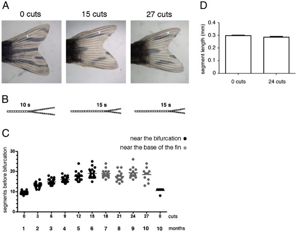
Zebrafish has emerged as a powerful model organism to study the process of regeneration. This teleost fish has the ability to regenerate various tissues and organs like the heart, spinal cord, retina and fins. In this study, we took advantage of the existence of an excellent morphological reference in the zebrafish caudal fin, the bony ray bifurcations, as a model to study positional information upon amputation. We investigated the existence of positional information for bifurcation formation by performing repeated amputations at different proximal-distal places along the fin.

RESULTS

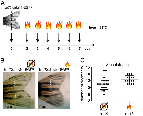
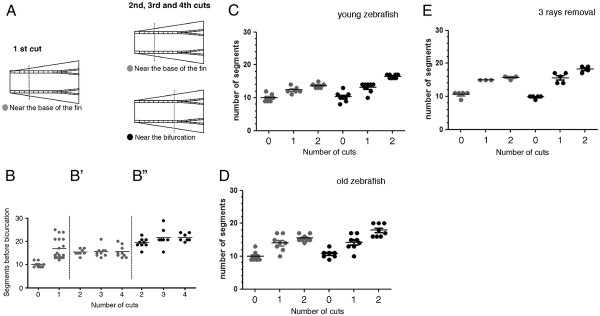
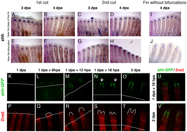
We show that, while amputations performed at a long distance from the bifurcation do not change its final proximal-distal position in the regenerated fin, consecutive amputations done at 1 segment proximal to the bifurcation (near the bifurcation) induce a positional reset and progressively shift its position distally. Furthermore, we investigated the potential role of Shh and Fgf signalling pathways in the determination of the bifurcation position and observed that they do not seem to be involved in this process.

CONCLUSIONS

Our results reveal that, an amputation near the bifurcation inhibits the formation of the regenerated bifurcation in the pre-amputation position, inducing a distalization of this structure. This shows that the positional memory for bony ray bifurcations depends on the proximal-distal level of the amputation.

摘要

背景

斑马鱼已成为研究再生过程的一种强大的模式生物。这种硬骨鱼能够再生各种组织和器官,如心脏、脊髓、视网膜和鳍。在本研究中,我们利用斑马鱼尾鳍中存在的一个出色的形态学参考——骨射线分支,作为研究截肢后位置信息的模型。我们通过在鳍的不同近端 - 远端位置进行重复截肢,研究了分支形成的位置信息的存在情况。

结果

我们发现,虽然在距离分支较远的位置进行截肢不会改变其在再生鳍中最终的近端 - 远端位置,但在分支近端 1 节处(靠近分支)连续进行截肢会引发位置重置,并使其位置逐渐向远端移动。此外,我们研究了 Sonic hedgehog(Shh)和成纤维细胞生长因子(Fgf)信号通路在确定分支位置中的潜在作用,发现它们似乎不参与这一过程。

结论

我们的结果表明,在分支附近进行截肢会抑制截肢前位置再生分支的形成,导致该结构向远端化。这表明骨射线分支的位置记忆取决于截肢的近端 - 远端水平。

https://cdn.ncbi.nlm.nih.gov/pmc/blobs/63f5/3484062/ef72d1e8805a/1471-213X-12-24-1.jpg

相似文献

2
Divergent requirements for fibroblast growth factor signaling in zebrafish maxillary barbel and caudal fin regeneration.
Dev Growth Differ. 2013 Feb;55(2):282-300. doi: 10.1111/dgd.12035. Epub 2013 Jan 28.
3
Widening control of fin inter-rays in zebrafish and inferences about actinopterygian fins.
J Anat. 2018 May;232(5):783-805. doi: 10.1111/joa.12785. Epub 2018 Feb 14.
6
The regenerative capacity of the zebrafish caudal fin is not affected by repeated amputations.
PLoS One. 2011;6(7):e22820. doi: 10.1371/journal.pone.0022820. Epub 2011 Jul 28.
7
Regrowth of zebrafish caudal fin regeneration is determined by the amputated length.
Sci Rep. 2020 Jan 20;10(1):649. doi: 10.1038/s41598-020-57533-6.
8
Tissue regeneration after injury in adult zebrafish: the regenerative potential of the caudal fin.
Dev Dyn. 2011 May;240(5):1271-7. doi: 10.1002/dvdy.22603. Epub 2011 Mar 15.
10
Robust and local positional information within a fin ray directs fin length during zebrafish regeneration.
Dev Growth Differ. 2018 Aug;60(6):354-364. doi: 10.1111/dgd.12558. Epub 2018 Jul 10.

引用本文的文献

1
Decaying and expanding Erk gradients process memory of skeletal size during zebrafish fin regeneration.
bioRxiv. 2025 Jan 23:2025.01.23.634576. doi: 10.1101/2025.01.23.634576.
5
Thyroid hormone regulates proximodistal patterning in fin rays.
Proc Natl Acad Sci U S A. 2023 May 23;120(21):e2219770120. doi: 10.1073/pnas.2219770120. Epub 2023 May 15.
6
Fin ray branching is defined by TRAP osteolytic tubules in zebrafish.
Proc Natl Acad Sci U S A. 2022 Nov 29;119(48):e2209231119. doi: 10.1073/pnas.2209231119. Epub 2022 Nov 23.
7
Genetically engineered zebrafish as models of skeletal development and regeneration.
Bone. 2023 Feb;167:116611. doi: 10.1016/j.bone.2022.116611. Epub 2022 Nov 14.
8
Crustacean leg regeneration restores complex microanatomy and cell diversity.
Sci Adv. 2022 Aug 26;8(34):eabn9823. doi: 10.1126/sciadv.abn9823. Epub 2022 Aug 24.
9
Common themes in tetrapod appendage regeneration: a cellular perspective.
Evodevo. 2019 Jun 17;10:11. doi: 10.1186/s13227-019-0124-7. eCollection 2019.
10
Identification of regenerative roadblocks via repeat deployment of limb regeneration in axolotls.
NPJ Regen Med. 2017 Nov 6;2:30. doi: 10.1038/s41536-017-0034-z. eCollection 2017.

本文引用的文献

1
Morphogen-based simulation model of ray growth and joint patterning during fin development and regeneration.
Development. 2012 Mar;139(6):1188-97. doi: 10.1242/dev.073452. Epub 2012 Feb 8.
3
The regenerative capacity of the zebrafish caudal fin is not affected by repeated amputations.
PLoS One. 2011;6(7):e22820. doi: 10.1371/journal.pone.0022820. Epub 2011 Jul 28.
4
Conserved mechanisms regulate outgrowth in zebrafish fins.
Nat Chem Biol. 2007 Oct;3(10):613-8. doi: 10.1038/nchembio.2007.36.
5
Distinct Wnt signaling pathways have opposing roles in appendage regeneration.
Development. 2007 Feb;134(3):479-89. doi: 10.1242/dev.001123. Epub 2006 Dec 21.
6
fgf20 is essential for initiating zebrafish fin regeneration.
Science. 2005 Dec 23;310(5756):1957-60. doi: 10.1126/science.1117637.
7
Fgf signaling instructs position-dependent growth rate during zebrafish fin regeneration.
Development. 2005 Dec;132(23):5173-83. doi: 10.1242/dev.02101. Epub 2005 Oct 26.
9
Tales of regeneration in zebrafish.
Dev Dyn. 2003 Feb;226(2):202-10. doi: 10.1002/dvdy.10220.
10
Ray-interray interactions during fin regeneration of Danio rerio.
Dev Biol. 2002 Dec 15;252(2):214-24. doi: 10.1006/dbio.2002.0848.

文献AI研究员

20分钟写一篇综述,助力文献阅读效率提升50倍。

立即体验

用中文搜PubMed

大模型驱动的PubMed中文搜索引擎

马上搜索

文档翻译

学术文献翻译模型,支持多种主流文档格式。

立即体验