• 文献检索
  • 文档翻译
  • 深度研究
  • 学术资讯
  • Suppr Zotero 插件Zotero 插件
  • 邀请有礼
  • 套餐&价格
  • 历史记录
应用&插件
Suppr Zotero 插件Zotero 插件浏览器插件Mac 客户端Windows 客户端微信小程序
定价
高级版会员购买积分包购买API积分包
服务
文献检索文档翻译深度研究API 文档MCP 服务
关于我们
关于 Suppr公司介绍联系我们用户协议隐私条款
关注我们

Suppr 超能文献

核心技术专利:CN118964589B侵权必究
粤ICP备2023148730 号-1Suppr @ 2026

文献检索

告别复杂PubMed语法,用中文像聊天一样搜索,搜遍4000万医学文献。AI智能推荐,让科研检索更轻松。

立即免费搜索

文件翻译

保留排版,准确专业,支持PDF/Word/PPT等文件格式,支持 12+语言互译。

免费翻译文档

深度研究

AI帮你快速写综述,25分钟生成高质量综述,智能提取关键信息,辅助科研写作。

立即免费体验

肿瘤性胃病变及其类似物:影像学表现谱。

Neoplastic stomach lesions and their mimickers: spectrum of imaging manifestations.

机构信息

The Ottawa Hospital, Ottawa, Ontario, Canada.

出版信息

Cancer Imaging. 2012 Aug 10;12(1):269-78. doi: 10.1102/1470-7330.2012.0031.

DOI:10.1102/1470-7330.2012.0031
PMID:22935192
原文链接:https://pmc.ncbi.nlm.nih.gov/articles/PMC3458788/
Abstract

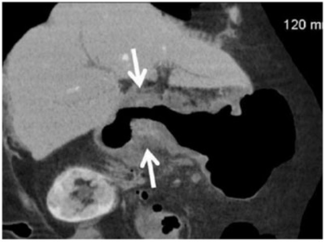
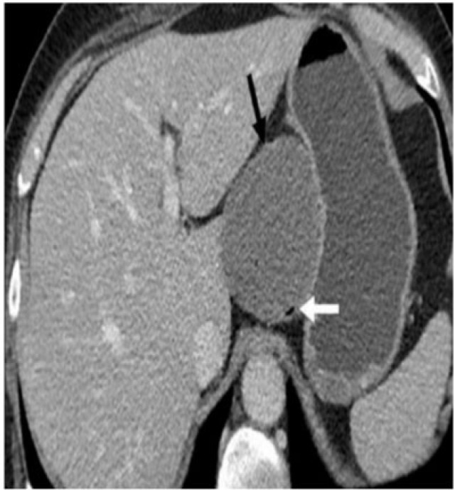
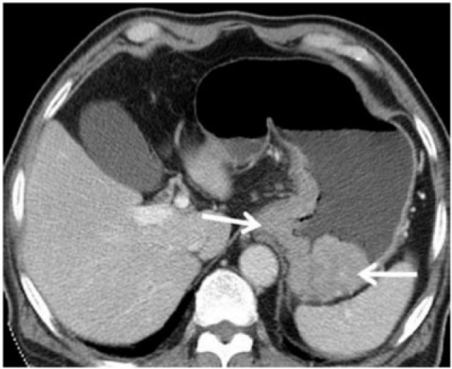
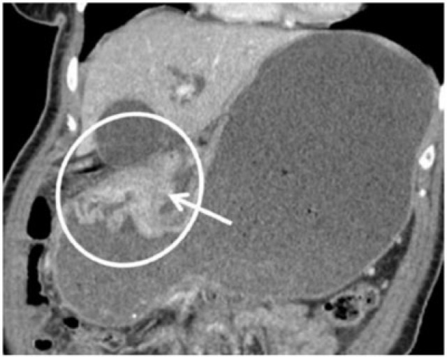
This review illustrates a wide spectrum of gastric neoplasms with emphasis on imaging findings helpful in characterizing various gastric neoplasms. Both the malignant and benign neoplasms along with focal gastric masses mimicking tumour are illustrated. Moreover, imaging clues to reach an accurate diagnosis are emphasized.

摘要

这篇综述阐述了广泛的胃肿瘤,重点介绍了有助于确定各种胃肿瘤特征的影像学表现。既阐述了恶性和良性肿瘤,也阐述了模拟肿瘤的局灶性胃肿块。此外,还强调了有助于准确诊断的影像学线索。

https://cdn.ncbi.nlm.nih.gov/pmc/blobs/7a1a/3458788/88b7582832c1/ci12003123.jpg
https://cdn.ncbi.nlm.nih.gov/pmc/blobs/7a1a/3458788/bd9cb4da9290/ci12003101.jpg
https://cdn.ncbi.nlm.nih.gov/pmc/blobs/7a1a/3458788/e7a7a21d5ced/ci12003102.jpg
https://cdn.ncbi.nlm.nih.gov/pmc/blobs/7a1a/3458788/827c5466f84a/ci12003103.jpg
https://cdn.ncbi.nlm.nih.gov/pmc/blobs/7a1a/3458788/a19cf38a5947/ci12003104.jpg
https://cdn.ncbi.nlm.nih.gov/pmc/blobs/7a1a/3458788/7aafe186e8ce/ci12003105.jpg
https://cdn.ncbi.nlm.nih.gov/pmc/blobs/7a1a/3458788/d6c2f1901d28/ci12003106.jpg
https://cdn.ncbi.nlm.nih.gov/pmc/blobs/7a1a/3458788/d1872409cdf8/ci12003107.jpg
https://cdn.ncbi.nlm.nih.gov/pmc/blobs/7a1a/3458788/669288619b74/ci12003108.jpg
https://cdn.ncbi.nlm.nih.gov/pmc/blobs/7a1a/3458788/7a39d6e19a7b/ci12003109.jpg
https://cdn.ncbi.nlm.nih.gov/pmc/blobs/7a1a/3458788/875ca5ffaf1a/ci12003110.jpg
https://cdn.ncbi.nlm.nih.gov/pmc/blobs/7a1a/3458788/60c6d3930b58/ci12003111.jpg
https://cdn.ncbi.nlm.nih.gov/pmc/blobs/7a1a/3458788/c9f1e75571fc/ci12003112.jpg
https://cdn.ncbi.nlm.nih.gov/pmc/blobs/7a1a/3458788/ff0f3e6ab22c/ci12003113.jpg
https://cdn.ncbi.nlm.nih.gov/pmc/blobs/7a1a/3458788/47a5f55e699e/ci12003114.jpg
https://cdn.ncbi.nlm.nih.gov/pmc/blobs/7a1a/3458788/b4d2047cd031/ci12003115.jpg
https://cdn.ncbi.nlm.nih.gov/pmc/blobs/7a1a/3458788/04e30c4cf6b9/ci12003116.jpg
https://cdn.ncbi.nlm.nih.gov/pmc/blobs/7a1a/3458788/4a56fe55663f/ci12003117.jpg
https://cdn.ncbi.nlm.nih.gov/pmc/blobs/7a1a/3458788/c2fcb367716e/ci12003118.jpg
https://cdn.ncbi.nlm.nih.gov/pmc/blobs/7a1a/3458788/f8a4b93fc2b9/ci12003119.jpg
https://cdn.ncbi.nlm.nih.gov/pmc/blobs/7a1a/3458788/76707641bb34/ci12003120.jpg
https://cdn.ncbi.nlm.nih.gov/pmc/blobs/7a1a/3458788/28ad39ca987f/ci12003121.jpg
https://cdn.ncbi.nlm.nih.gov/pmc/blobs/7a1a/3458788/a646651f9f4f/ci12003122.jpg
https://cdn.ncbi.nlm.nih.gov/pmc/blobs/7a1a/3458788/88b7582832c1/ci12003123.jpg

相似文献

1
Neoplastic stomach lesions and their mimickers: spectrum of imaging manifestations.肿瘤性胃病变及其类似物:影像学表现谱。
Cancer Imaging. 2012 Aug 10;12(1):269-78. doi: 10.1102/1470-7330.2012.0031.
2
Duodenal tumors on cross-sectional imaging with emphasis on multidetector computed tomography: a pictorial review.[文章标题]:多排螺旋 CT 对十二指肠肿瘤的影像学诊断:图像评价 **注意**:这是一个示例回答,你可以根据实际提供的文本进行翻译。
Diagn Interv Radiol. 2020 May;26(3):193-199. doi: 10.5152/dir.2019.19241.
3
Gastric Lipoma: A Review of the Literature.胃脂肪瘤:文献综述
Ultrasound Q. 2018 Sep;34(3):119-121. doi: 10.1097/RUQ.0000000000000338.
4
Gastric inflammatory fibroid polyp mimicking a gastrointestinal stromal tumour.酷似胃肠道间质瘤的胃炎性纤维性息肉
Rev Esp Enferm Dig. 2016 Aug;108(8):497-8.
5
Contrast-enhanced computed tomography may be helpful for characterizing and staging canine gastric tumors.对比增强计算机断层扫描可能有助于对犬胃肿瘤进行特征描述和分期。
Vet Radiol Ultrasound. 2019 Jan;60(1):7-18. doi: 10.1111/vru.12677. Epub 2018 Aug 19.
6
Unusual gastric tumors and tumor-like lesions: Radiological with pathological correlation and literature review.特殊胃肿瘤及肿瘤样病变:影像学与病理对照及文献综述
World J Gastroenterol. 2017 Apr 14;23(14):2493-2504. doi: 10.3748/wjg.v23.i14.2493.
7
Computed tomography of gastrocolic ligament: involvement in malignant tumors of the stomach.胃结肠韧带的计算机断层扫描:与胃癌的关系
Abdom Imaging. 2007 Jan-Feb;32(1):59-65. doi: 10.1007/s00261-006-9000-8.
8
Gastric manifestations of autoimmune deficiency syndrome-related Kaposi's sarcoma on computed tomography.计算机断层扫描显示的自身免疫缺陷综合征相关卡波西肉瘤的胃部表现
J Comput Tomogr. 1986 Jan;10(1):85-8. doi: 10.1016/0149-936x(86)90018-4.
9
Gastric manifestations of AIDS: radiographic findings on upper gastrointestinal examination.艾滋病的胃部表现:上消化道检查的影像学结果
Gastrointest Radiol. 1991 Spring;16(2):95-8. doi: 10.1007/BF01887318.
10
[Benign gastric tumors: anatomo-radioclinical data].[胃良性肿瘤:解剖放射临床资料]
J Radiol Electrol Med Nucl. 1969 Mar-Apr;50(3):220-2.

引用本文的文献

1
Delayed Metastatic Cholangiocarcinoma to the Stomach With Imaging Characteristics Resembling Gastrointestinal Stromal Tumor: A Case Report and Review of the Literature.具有类似胃肠道间质瘤影像学特征的延迟性胃转移胆管癌:一例报告及文献复习
Cureus. 2023 Jul 12;15(7):e41785. doi: 10.7759/cureus.41785. eCollection 2023 Jul.
2
Gastrointestinal malignancies in pregnancy.妊娠期胃肠道恶性肿瘤
Abdom Radiol (NY). 2023 May;48(5):1709-1723. doi: 10.1007/s00261-022-03788-8. Epub 2023 Jan 6.
3
Identification of a Five-Gene Panel to Assess Prognosis for Gastric Cancer.

本文引用的文献

1
An unusual case of recurrent gastric abscess.复发性胃脓肿一例罕见病例。
Gastroenterol Hepatol (N Y). 2008 Sep;4(9):641-3.
2
Imaging of gastroenteropancreatic neuroendocrine tumors.胃肠胰神经内分泌肿瘤的影像学检查
World J Clin Oncol. 2011 Jan 10;2(1):28-43. doi: 10.5306/wjco.v2.i1.28.
3
Characteristics of ectopic pancreas in dynamic gadolinium-enhanced MRI.动态钆增强磁共振成像中异位胰腺的特征
鉴定一个五基因标志物panel 用于评估胃癌预后。
Biomed Res Int. 2022 Feb 9;2022:5593619. doi: 10.1155/2022/5593619. eCollection 2022.
4
Imaging of Kaposi sarcoma.卡波西肉瘤的影像学表现。
Abdom Radiol (NY). 2021 Nov;46(11):5297-5306. doi: 10.1007/s00261-021-03205-6. Epub 2021 Jul 13.
5
Sarcomatoid carcinoma of the pancreas - multimodality imaging findings with serial imaging follow-up: A case report and review of literature.胰腺肉瘤样癌——多模态影像学表现及系列影像学随访:一例病例报告并文献复习
World J Clin Cases. 2021 May 6;9(13):3102-3113. doi: 10.12998/wjcc.v9.i13.3102.
6
The impact of age on prognosis in patients with gastric cancer: experience in a tertiary care centre.年龄对胃癌患者预后的影响:三级医疗中心的经验
J Gastrointest Oncol. 2020 Dec;11(6):1233-1241. doi: 10.21037/jgo-20-139.
7
Gastric Cancer Presenting as Isolated Ascites: A Diagnostic Challenge.以孤立性腹水为表现的胃癌:一项诊断挑战。
Eur J Case Rep Intern Med. 2019 Jun 21;6(7):001141. doi: 10.12890/2019_001141. eCollection 2019.
8
MDCT imaging of the stomach: advances and applications.胃的多层螺旋CT成像:进展与应用
Br J Radiol. 2017 Jan;90(1069):20160412. doi: 10.1259/bjr.20160412. Epub 2016 Dec 8.
9
Gastric lipoma: an unusual cause of dyspeptic symptoms.胃脂肪瘤:消化不良症状的不寻常病因。
BMJ Case Rep. 2016 Jun 22;2016:bcr2016215297. doi: 10.1136/bcr-2016-215297.
10
Visceral Kaposi's Sarcoma Presenting as Upper Gastrointestinal Bleeding.以消化道出血为表现的内脏型卡波西肉瘤
Case Rep Gastrointest Med. 2015;2015:438973. doi: 10.1155/2015/438973. Epub 2015 May 6.
Abdom Imaging. 2010 Feb;35(1):85-7. doi: 10.1007/s00261-008-9491-6. Epub 2008 Dec 2.
4
Primary gastrointestinal lymphoma: spectrum of imaging findings with pathologic correlation.原发性胃肠道淋巴瘤:影像表现谱与病理对照
Radiographics. 2007 Sep-Oct;27(5):1371-88. doi: 10.1148/rg.275065151.
5
From the archives of the AFIP: Gastrointestinal carcinoids: imaging features with clinicopathologic comparison.源自武装部队病理研究所档案:胃肠道类癌:影像学特征与临床病理对照
Radiographics. 2007 Jan-Feb;27(1):237-57. doi: 10.1148/rg.271065169.
6
Gastrointestinal stromal tumor: role of CT in diagnosis and in response evaluation and surveillance after treatment with imatinib.胃肠道间质瘤:CT在诊断、伊马替尼治疗后疗效评估及监测中的作用
Radiographics. 2006 Mar-Apr;26(2):481-95. doi: 10.1148/rg.262055097.
7
Gastrointestinal stromal tumors of the stomach: CT findings and prediction of malignancy.胃胃肠道间质瘤:CT表现及恶性预测
AJR Am J Roentgenol. 2004 Oct;183(4):893-8. doi: 10.2214/ajr.183.4.1830893.
8
Preoperative staging of gastric adenocarcinoma: comparison of helical CT and endoscopic US.胃腺癌的术前分期:螺旋CT与内镜超声的比较
Radiology. 2004 Feb;230(2):465-71. doi: 10.1148/radiol.2302020828.
9
Wall thickening of the gastric antrum as a normal finding: multidetector CT with cadaveric comparison.胃窦壁增厚作为正常表现:多排螺旋CT与尸体解剖对照研究
AJR Am J Roentgenol. 2003 Oct;181(4):973-9. doi: 10.2214/ajr.181.4.1810973.
10
Benign and malignant lesions of the stomach: evaluation of CT criteria for differentiation.胃的良性与恶性病变:CT鉴别标准的评估
Radiology. 2003 Jul;228(1):166-71. doi: 10.1148/radiol.2281020623. Epub 2003 May 20.