JHU ICMIC Program, Division of Cancer Imaging Research, The Russell H Morgan Department of Radiology and Radiological Science, Johns Hopkins University School of Medicine, Baltimore, Maryland, United States of America.
PLoS One. 2012;7(8):e44078. doi: 10.1371/journal.pone.0044078. Epub 2012 Aug 28.
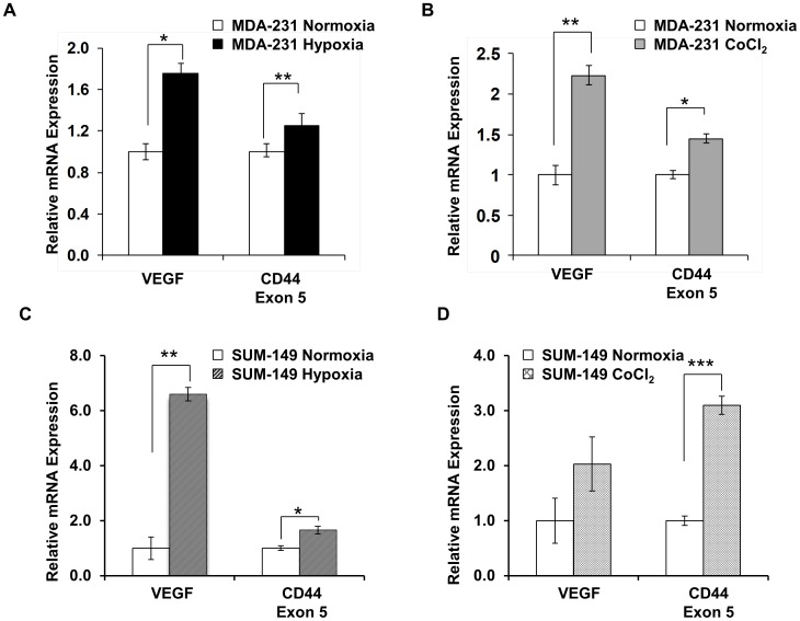
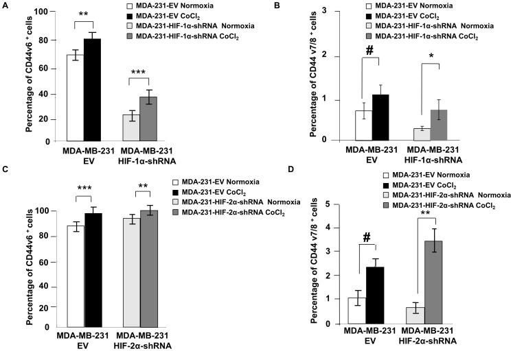
The CD44 transmembrane glycoproteins play multifaceted roles in tumor progression and metastasis. CD44 expression has also been associated with stem-like breast cancer cells. Hypoxia commonly occurs in tumors and is a major cause of radiation and chemo-resistance. Hypoxia is known to inhibit differentiation and facilitates invasion and metastasis. Here we have investigated the effect of hypoxia on CD44 and two of its isoforms in MDA-MB-231 and SUM-149 triple negative human breast cancer cells and MDA-MB-231 tumors using imaging and molecular characterization.
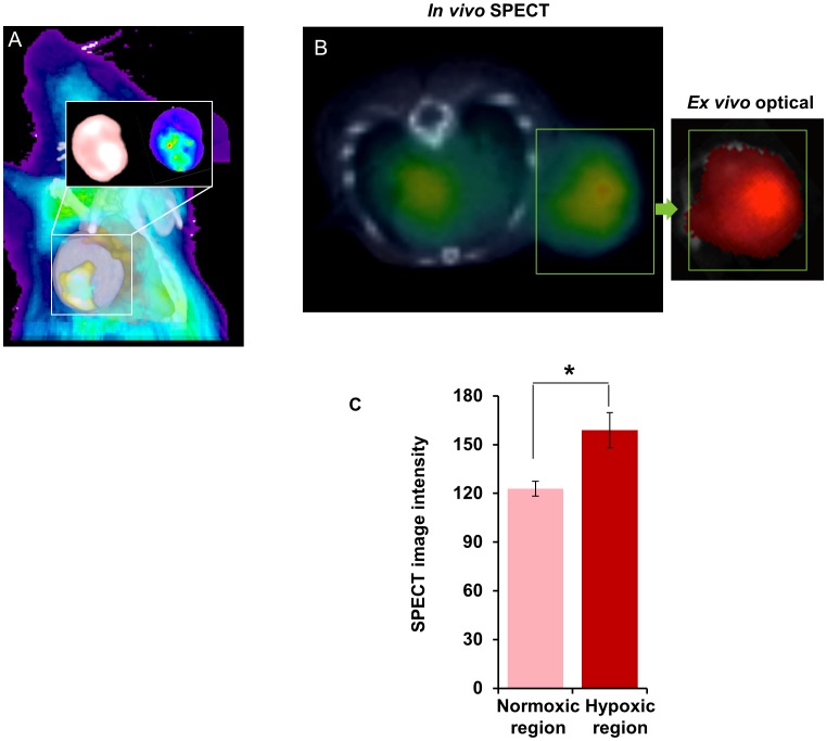
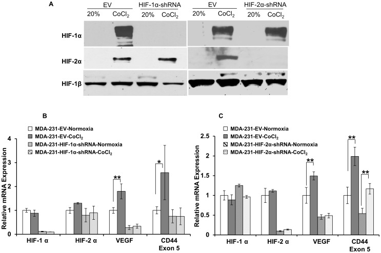
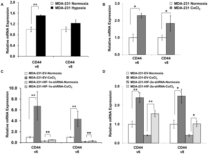
The roles of hypoxia and hypoxia inducible factor (HIF) in regulating the expression of CD44 and its variant isoforms (CD44v6, CD44v7/8) were investigated in human breast cancer cells, by quantitative real-time polymerase chain reaction (qRT-PCR) to determine mRNA levels, and fluorescence associated cell sorting (FACS) to determine cell surface expression of CD44, under normoxic and hypoxic conditions. In vivo imaging studies with tumor xenografts derived from MDA-MD-231 cells engineered to express tdTomato red fluorescence protein under regulation of hypoxia response elements identified co-localization between hypoxic fluorescent regions and increased concentration of (125)I-radiolabeled CD44 antibody.
Our data identified HIF-1α as a regulator of CD44 that increased the number of CD44 molecules and the percentage of CD44 positive cells expressing variant exons v6 and v7/8 in breast cancer cells under hypoxic conditions. Data from these cell studies were further supported by in vivo observations that hypoxic tumor regions contained cells with a higher concentration of CD44 expression.
CD44 跨膜糖蛋白在肿瘤的进展和转移中发挥着多方面的作用。CD44 的表达也与乳腺癌干细胞有关。缺氧通常发生在肿瘤中,是导致放射和化疗耐药的主要原因。已知缺氧抑制分化,促进侵袭和转移。在这里,我们通过成像和分子特征研究了缺氧对 MDA-MB-231 和 SUM-149 三阴性人乳腺癌细胞和 MDA-MB-231 肿瘤中 CD44 及其两种同工型(CD44v6、CD44v7/8)的影响。
通过定量实时聚合酶链反应(qRT-PCR)确定 mRNA 水平,通过荧光相关细胞分选(FACS)确定 CD44 细胞表面表达,研究了缺氧和缺氧诱导因子(HIF)在调节人乳腺癌细胞中 CD44 及其变体同工型(CD44v6、CD44v7/8)表达中的作用,在常氧和缺氧条件下。利用由 MDA-MD-231 细胞衍生的肿瘤异种移植进行体内成像研究,这些细胞经过工程改造后,在缺氧反应元件的调控下表达 tdTomato 红色荧光蛋白,鉴定出缺氧荧光区域与(125)I 放射性标记 CD44 抗体浓度增加的共定位。
我们的数据确定 HIF-1α 是 CD44 的调节剂,在缺氧条件下增加了乳腺癌细胞中 CD44 分子的数量和表达变体外显子 v6 和 v7/8 的 CD44 阳性细胞的百分比。这些细胞研究的数据进一步得到了体内观察的支持,即缺氧肿瘤区域含有 CD44 表达浓度更高的细胞。