Mirrione Martine M, Tsirka Stella E
Department of Pharmacological Sciences, Molecular and Cellular Pharmacology, Stony Brook University, Stony Brook, NY 11794-8651, USA.
Epilepsy Res Treat. 2011;2011:369295. doi: 10.1155/2011/369295. Epub 2011 Jul 7.
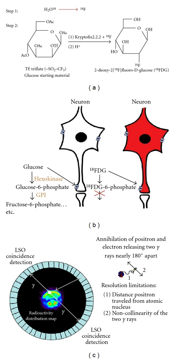
Small animal neuroimaging has become increasingly available to researchers, expanding the breadth of questions studied with these methods. Applying these noninvasive techniques to the open questions underlying epileptogenesis is no exception. A major advantage of small animal neuroimaging is its translational appeal. Studies can be well controlled and manipulated, examining the living brain in the animal before, during, and after the disease onset or disease treatment. The results can also be compared to data collected on human patients. Over the past decade, we and others have explored metabolic patterns in animal models of epilepsy to gain insight into the circuitry underlying development of the disease. In this paper, we provide technical details on how metabolic imaging that uses 2-deoxy-2[(18)F]fluoro-D-glucose ((18)FDG) and positron emission tomography (PET) is performed and explain the strengths and limitations of these studies. We will also highlight recent advances toward understanding epileptogenesis through small animal imaging.
小动物神经成像技术已越来越多地为研究人员所用,拓宽了使用这些方法所研究问题的广度。将这些非侵入性技术应用于癫痫发生的相关开放性问题也不例外。小动物神经成像的一个主要优势在于其转化应用价值。研究可以得到很好的控制和操作,能够在疾病发作前、发作期间以及疾病治疗后对动物的活体大脑进行检查。研究结果还可以与人类患者收集的数据进行比较。在过去十年中,我们和其他研究人员探索了癫痫动物模型中的代谢模式,以深入了解该疾病发展背后的神经回路。在本文中,我们提供了关于如何进行使用2-脱氧-2-[(18)F]氟-D-葡萄糖((18)FDG)和正电子发射断层扫描(PET)的代谢成像的技术细节,并解释这些研究的优势和局限性。我们还将重点介绍通过小动物成像在理解癫痫发生方面的最新进展。