Department of Physics and Astronomy, University of North Carolina, Chapel Hill, NC 27599, USA.
Phys Med Biol. 2012 Sep 21;57(18):5749-63. doi: 10.1088/0031-9155/57/18/5749. Epub 2012 Sep 5.
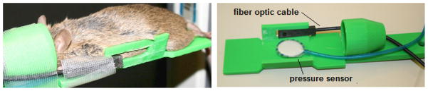
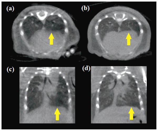
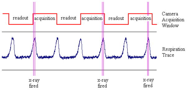
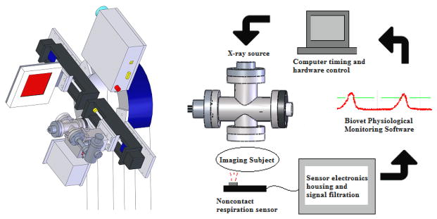
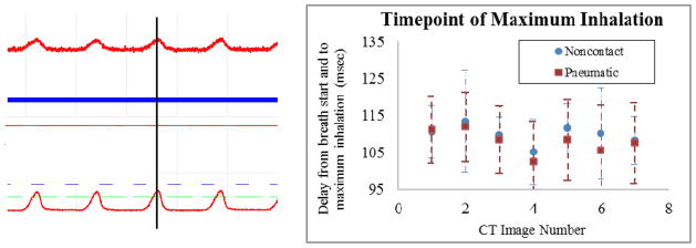
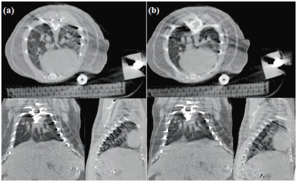
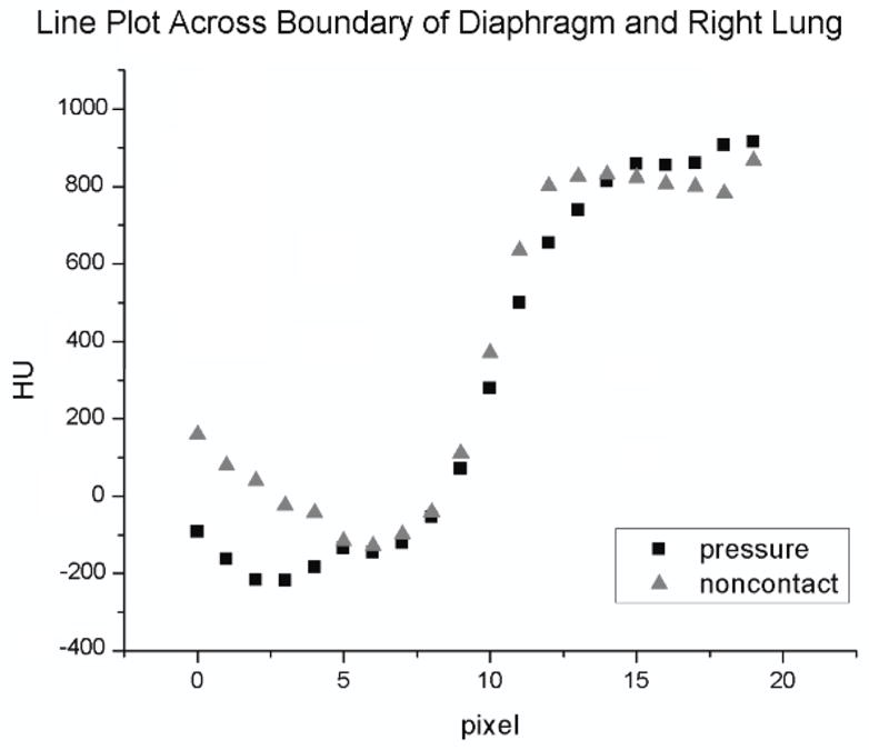
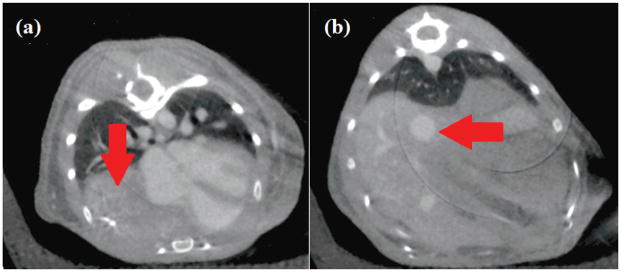
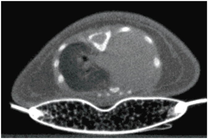
A cone beam micro-CT has previously been utilized along with a pressure-tracking respiration sensor to acquire prospectively gated images of both wild-type mice and various adult murine disease models. While the pressure applied to the abdomen of the subject by this sensor is small and is generally without physiological effect, certain disease models of interest, as well as very young animals, are prone to atelectasis with added pressure, or they generate too weak a respiration signal with this method to achieve optimal prospective gating. In this work we present a new fibre-optic displacement sensor which monitors respiratory motion of a subject without requiring physical contact. The sensor outputs an analogue signal which can be used for prospective respiration gating in micro-CT imaging. The device was characterized and compared against a pneumatic air chamber pressure sensor for the imaging of adult wild-type mice. The resulting images were found to be of similar quality with respect to physiological motion blur; the quality of the respiration signal trace obtained using the non-contact sensor was comparable to that of the pressure sensor and was superior for gating purposes due to its better signal-to-noise ratio. The non-contact sensor was then used to acquire in-vivo micro-CT images of a murine model for congenital diaphragmatic hernia and of 11-day-old mouse pups. In both cases, quality CT images were successfully acquired using this new respiration sensor. Despite the presence of beam hardening artefacts arising from the presence of a fibre-optic cable in the imaging field, we believe this new technique for respiration monitoring and gating presents an opportunity for in-vivo imaging of disease models which were previously considered too delicate for established animal handling methods.
先前已经使用锥形束微 CT 结合压力跟踪呼吸传感器来获取野生型小鼠和各种成年小鼠疾病模型的前瞻性门控图像。虽然该传感器施加在受试动物腹部的压力很小,通常没有生理影响,但某些感兴趣的疾病模型以及非常年幼的动物,在增加压力时容易发生肺不张,或者它们使用这种方法产生的呼吸信号太弱,无法实现最佳的前瞻性门控。在这项工作中,我们提出了一种新的光纤位移传感器,它可以在不接触受试动物的情况下监测呼吸运动。传感器输出模拟信号,可用于微 CT 成像中的前瞻性呼吸门控。对该设备进行了表征,并与气动空气室压力传感器进行了比较,用于成年野生型小鼠的成像。结果表明,两种传感器获得的图像在生理运动模糊方面具有相似的质量;使用非接触式传感器获得的呼吸信号迹线的质量与压力传感器相当,并且由于其信噪比更高,因此在门控方面更优。然后,使用非接触式传感器成功地对先天性膈疝的小鼠模型和 11 天大的小鼠幼崽进行了体内 micro-CT 成像。在这两种情况下,都成功地使用这种新的呼吸传感器获得了高质量的 CT 图像。尽管在成像场中存在光纤电缆会产生束硬化伪影,但我们相信这种用于呼吸监测和门控的新技术为以前认为由于现有的动物处理方法太脆弱而无法进行的疾病模型的体内成像提供了机会。