Universität Bayreuth, Lehrstuhl Biopolymere, Universitätsstr, 30, D-95447 Bayreuth, Germany.
Retrovirology. 2012 Sep 10;9:73. doi: 10.1186/1742-4690-9-73.
The ribonuclease H (RNase H) domains of retroviral reverse transcriptases play an essential role in the replication cycle of retroviruses. During reverse transcription of the viral genomic RNA, an RNA/DNA hybrid is created whose RNA strand needs to be hydrolyzed by the RNase H to enable synthesis of the second DNA strand by the DNA polymerase function of the reverse transcriptase. Here, we report the solution structure of the separately purified RNase H domain from prototype foamy virus (PFV) revealing the so-called C-helix and the adjacent basic loop, which both were suggested to be important in substrate binding and activity.
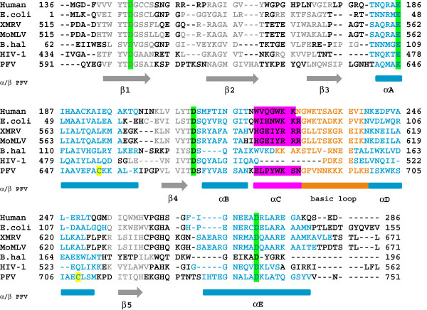
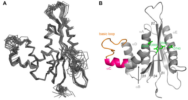
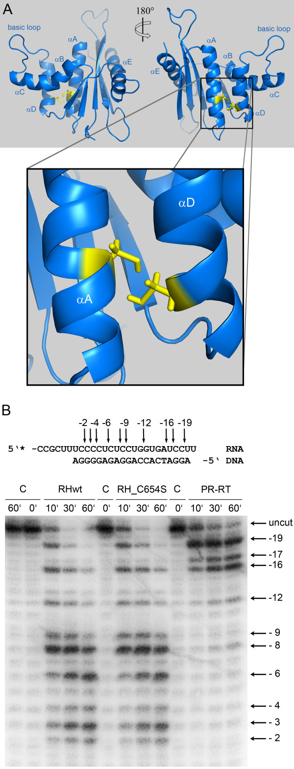
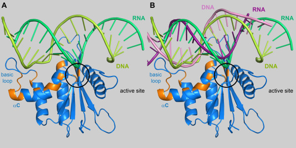
The solution structure of PFV RNase H shows that it contains a mixed five-stranded β-sheet, which is sandwiched by four α-helices (A-D), including the C-helix, on one side and one α-helix (helix E) on the opposite side. NMR titration experiments demonstrate that upon substrate addition signal changes can be detected predominantly in the basic loop as well as in the C-helix. All these regions are oriented towards the bound substrate. In addition, signal intensities corresponding to residues in the B-helix and the active site decrease, while only minor or no changes of the overall structure of the RNase H are detectable upon substrate binding. Dynamic studies confirm the monomeric state of the RNase H domain. Structure comparisons with HIV-1 RNase H, which lacks the basic protrusion, indicate that the basic loop is relevant for substrate interaction, while the C-helix appears to fulfill mainly structural functions, i.e. positioning the basic loop in the correct orientation for substrate binding.
The structural data of PFV RNase H demonstrate the importance of the basic loop, which contains four positively charged lysines, in substrate binding and the function of the C-helix in positioning of the loop. In the dimeric full length HIV-1 RT, the function of the basic loop is carried out by a different loop, which also harbors basic residues, derived from the connection domain of the p66 subunit. Our results suggest that RNases H which are also active as separate domains might need a functional basic loop for proper substrate binding.
逆转录病毒反转录酶的核糖核酸酶 H(RNase H)结构域在逆转录病毒的复制周期中发挥着重要作用。在病毒基因组 RNA 的反转录过程中,会产生一个 RNA/DNA 杂交体,其 RNA 链需要由 RNase H 水解,以便反转录酶的 DNA 聚合酶功能合成第二条 DNA 链。在这里,我们报告了从原型泡沫病毒(PFV)中分离纯化的 RNase H 结构域的溶液结构,揭示了所谓的 C-螺旋和相邻的碱性环,这两者都被认为在底物结合和活性中很重要。
PFV RNase H 的溶液结构表明,它包含一个混合的五股β-折叠,由四个α-螺旋(A-D)夹在一侧,另一侧有一个α-螺旋(螺旋 E)。NMR 滴定实验表明,在底物加入后,信号变化主要可以在碱性环以及 C-螺旋中检测到。所有这些区域都朝向结合的底物。此外,在结合底物时,对应于 B-螺旋和活性位点的残基的信号强度降低,而仅检测到 RNase H 整体结构的微小或无变化。动态研究证实了 RNase H 结构域的单体状态。与缺乏碱性突出的 HIV-1 RNase H 的结构比较表明,碱性环与底物相互作用有关,而 C-螺旋似乎主要起结构作用,即正确定位碱性环以进行底物结合。
PFV RNase H 的结构数据表明,碱性环在底物结合和 C-螺旋在正确定位环方面的功能中具有重要意义,该环包含四个带正电荷的赖氨酸。在二聚体全长 HIV-1 RT 中,碱性环的功能由连接 p66 亚基的连接域衍生的另一个环承担,该环也含有碱性残基。我们的结果表明,作为独立结构域发挥作用的 RNase H 可能需要一个功能性的碱性环才能正确结合底物。