Muttarak M, Sriburi T
Department of Radiology, Chiang Mai University, Chiang Mai, Thailand.
Biomed Imaging Interv J. 2012 Jan;8(1):e7. doi: 10.2349/biij.8.1.e7. Epub 2012 Jan 1.
To document the types of congenital renal anomalies detected in adulthood, the clinical presentation and complications of these renal anomalies, and the most useful imaging modality in detecting a renal anomaly.
This study was approved by the institutional review board and informed consent was waived. Between January 2007 and January 2011, the clinical data and imaging studies of 28 patients older than 18 years diagnosed with renal anomaly at the authors' institution were retrospectively reviewed. Renal anomalies in this study included only those with abnormality in position and in form.
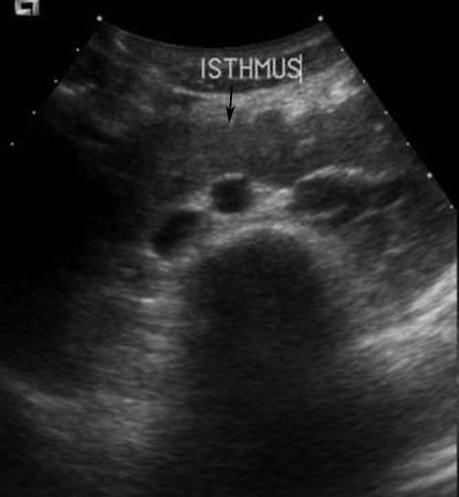
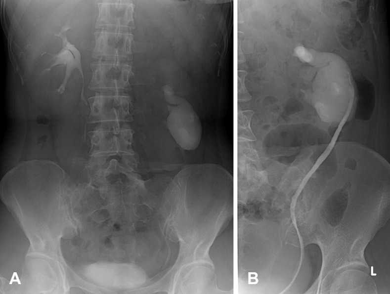
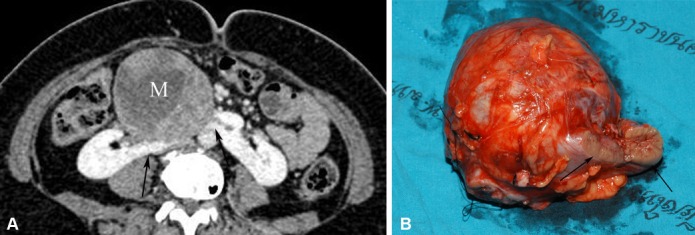
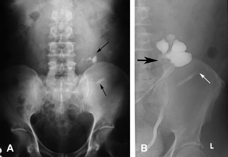
Of these 28 patients, 22 underwent imaging studies and their results constituted the material of this study. Of the 22 patients, 14 had horseshoe kidneys (HSK), four had crossed renal ectopia and four had malrotation. Sixteen patients were men and six were women. The patients ranged in age from 19 to 74 years (mean age 51.1 years). Clinical presentations were abdominal pain (13), fever (13), haematuria (4), palpable mass (2), asymptomatic (2), polyuria (1) dysuria (1), blurred vision (1), and headache with weakness of left extremities (1). Imaging studies included abdominal radiograph (15), intravenous pyelography (IVP) (8), retrograde pyelography (RP) (4), ultrasonography (US) (7), and computed tomography (CT) (9). Associated complications included urinary tract stones (17), urinary tract infection (16), hydronephrosis (12), and tumours (2). Abdominal radiograph suggested renal anomalies in nine out of 15 studies. IVP, RP, US and CT suggested anomalies in all patients who had these studies performed. However, CT was the best imaging modality to evaluate anatomy, function and complications of patients with renal anomalies.
HSK was the most common renal anomaly, with abdominal pain and fever being the most common presentations. UTI and stones were the most common complications. IVP, RP, US and CT can be used to diagnose renal anomalies but CT is the best imaging modality to evaluate renal anatomy, function and its complications.
记录成年期检测到的先天性肾异常类型、这些肾异常的临床表现和并发症,以及检测肾异常最有用的影像学检查方法。
本研究经机构审查委员会批准,无需知情同意。2007年1月至2011年1月期间,对作者所在机构诊断为肾异常的28例18岁以上患者的临床资料和影像学检查进行回顾性分析。本研究中的肾异常仅包括位置和形态异常的情况。
这28例患者中,22例接受了影像学检查,其结果构成了本研究的材料。在这22例患者中,14例为马蹄肾(HSK),4例为交叉异位肾,4例为旋转不良。16例为男性,6例为女性。患者年龄在19至74岁之间(平均年龄51.1岁)。临床表现为腹痛(13例)、发热(13例)、血尿(4例)、可触及肿块(2例)、无症状(2例)、多尿(1例)、排尿困难(1例)、视力模糊(1例)、头痛伴左侧肢体无力(1例)。影像学检查包括腹部X线平片(15例)、静脉肾盂造影(IVP)(8例)、逆行肾盂造影(RP)(4例)、超声检查(US)(7例)和计算机断层扫描(CT)(9例)。相关并发症包括尿路结石(17例)、尿路感染(16例)、肾积水(12例)和肿瘤(2例)。15例腹部X线平片检查中有9例提示肾异常。IVP、RP、US和CT对所有进行这些检查的患者均提示异常。然而,CT是评估肾异常患者的解剖结构、功能和并发症的最佳影像学检查方法。
马蹄肾是最常见的肾异常,腹痛和发热是最常见的表现。尿路感染和结石是最常见的并发症。IVP、RP、US和CT均可用于诊断肾异常,但CT是评估肾解剖结构、功能及其并发症的最佳影像学检查方法。