Lewis J C, Hantgan R R, Stevenson S C, Thornburg T, Kieffer N, Guichard J, Breton-Gorius J
Department of Pathology, Bowman Gray School of Medicine of Wake Forest University, Winston-Salem, North Carolina 27103.
Am J Pathol. 1990 Jan;136(1):239-52.
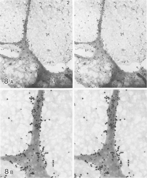
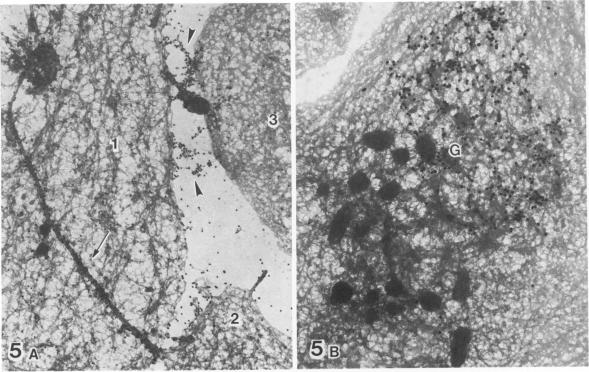
Platelet membrane glycoprotein IIb-IIIa plays a focal role in primary hemostasis by serving as the cell surface receptor for fibrinogen. Recent studies by several groups have suggested that GPIIb-IIIa, which is dispersed randomly in the resting cell, undergoes migration leading to receptor clustering after platelet activation. The authors have investigated this activation-dependent relocation of fibrinogen receptors on platelets adherent to a standardized artificial surface. The correlative use of immunogold electron microscopy, ligand-gold binding, and stereo (three-dimensional) electron microscopy (EM) revealed specific localization of fibrinogen and its receptor at points of platelet to platelet interaction. Fibrinogen distribution on the plasma membrane, studied through the use of fibrinogen-gold conjugates with whole-mount adherent platelets, was primarily over the granulomere and at the cell periphery corresponding to sites of platelet-platelet interaction. Compared with the general hyalomere, fibrinogen density over the granulomere and at contact regions was increased 12-fold and 22-fold, respectively, and the specificity of binding at these sites was verified by positive competition with native fibrinogen, one of its degradation products (Fragment D1), and by monoclonal antibodies (HP1-1d and AP-2) specific for GPIIb-IIIa. The distribution of receptor antigens, localized by immunogold EM using antibodies against GPIIb-IIIa, also was localized over the hyalomere, where fibrinogen did not bind. To understand this apparently nonfunctional hyalomere GPIIb-IIIa further, correlative immunocytochemistry was performed using polyclonal and monoclonal antibodies for GPIIb and GPIIIa simultaneously. Colocalization of the antigens was observed consistently over the granulomere and at regions of cell contact, whereas the hyalomere antigens tended to be nonassociated. The studies document GPIIb-IIIa as a function complex at sites of cell interaction where fibrinogen binds.
血小板膜糖蛋白IIb-IIIa作为纤维蛋白原的细胞表面受体,在初级止血过程中发挥着关键作用。多个研究小组最近的研究表明,在静息细胞中随机分布的GPIIb-IIIa,在血小板激活后会发生迁移,导致受体聚集。作者研究了粘附在标准化人工表面的血小板上纤维蛋白原受体这种依赖激活的重新定位。免疫金电子显微镜、配体-金结合和立体(三维)电子显微镜(EM)的相关应用揭示了纤维蛋白原及其受体在血小板与血小板相互作用点的特异性定位。通过使用纤维蛋白原-金缀合物与全量粘附血小板研究血浆膜上的纤维蛋白原分布,发现其主要位于颗粒区以及对应血小板-血小板相互作用部位的细胞周边。与一般的透明区相比,颗粒区和接触区域的纤维蛋白原密度分别增加了12倍和22倍,并且通过与天然纤维蛋白原、其降解产物之一(片段D1)的阳性竞争以及针对GPIIb-IIIa的单克隆抗体(HP1-1d和AP-2)验证了这些位点结合的特异性。使用针对GPIIb-IIIa的抗体通过免疫金EM定位的受体抗原分布,也位于透明区,而纤维蛋白原不在此结合。为了进一步了解这种明显无功能的透明区GPIIb-IIIa,同时使用针对GPIIb和GPIIIa的多克隆和单克隆抗体进行相关免疫细胞化学研究。在颗粒区和细胞接触区域始终观察到抗原的共定位,而透明区抗原往往不相关。这些研究证明GPIIb-IIIa是纤维蛋白原结合的细胞相互作用位点处的功能复合物。