Dept, of Neurology, University of Würzburg, Josef-Schneider-Str. 11, 97080, Würzburg, Germany.
BMC Neurol. 2012 Sep 13;12:92. doi: 10.1186/1471-2377-12-92.
Compensation of brain injury in multiple sclerosis (MS) may in part work through mechanisms involving neuronal plasticity on local and interregional scales. Mechanisms limiting excessive neuronal activity may have special significance for retention and (re-)acquisition of lost motor skills in brain injury. However, previous neurophysiological studies of plasticity in MS have investigated only excitability enhancing plasticity and results from neuroimaging are ambiguous. Thus, the aim of this study was to probe long-term depression-like central motor plasticity utilizing continuous theta-burst stimulation (cTBS), a non-invasive brain stimulation protocol. Because cTBS also may trigger behavioral effects through local interference with neuronal circuits, this approach also permitted investigating the functional role of the primary motor cortex (M1) in force control in patients with MS.
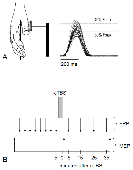
We used cTBS and force recordings to examine long-term depression-like central motor plasticity and behavioral consequences of a M1 lesion in 14 patients with stable mild-to-moderate MS (median EDSS 1.5, range 0 to 3.5) and 14 age-matched healthy controls. cTBS consisted of bursts (50 Hz) of three subthreshold biphasic magnetic stimuli repeated at 5 Hz for 40 s over the hand area of the left M1. Corticospinal excitability was probed via motor-evoked potentials (MEP) in the abductor pollicis brevis muscle over M1 before and after cTBS. Force production performance was assessed in an isometric right thumb abduction task by recording the number of hits into a predefined force window.
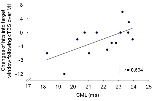
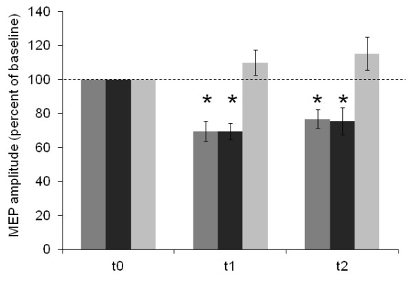
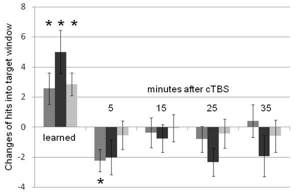
cTBS reduced MEP amplitudes in the contralateral abductor pollicis brevis muscle to a comparable extent in control subjects (69 ± 22% of baseline amplitude, p < 0.001) and in MS patients (69 ± 18%, p < 0.001). In contrast, post-cTBS force production performance was only impaired in controls (2.2 ± 2.8, p = 0.011), but not in MS patients (2.0 ± 4.4, p = 0.108). The decline in force production performance following cTBS correlated with corticomuscular latencies (CML) in MS patients, but did not correlate with MEP amplitude reduction in patients or controls.
Long-term depression-like plasticity remains largely intact in mild-to-moderate MS. Increasing brain injury may render the neuronal networks less responsive toward lesion-induction by cTBS.
多发性硬化症(MS)患者的脑损伤补偿部分通过局部和区域间尺度上涉及神经元可塑性的机制起作用。限制过度神经元活动的机制对于保留和(重新)获得脑损伤导致的运动技能丧失可能具有特殊意义。然而,之前关于 MS 中可塑性的神经生理学研究仅调查了兴奋性增强的可塑性,神经影像学的结果也存在矛盾。因此,本研究旨在利用连续 theta 爆发刺激(cTBS)探测类似长时程抑制的中枢运动可塑性,这是一种非侵入性的脑刺激方案。由于 cTBS 也可能通过局部干扰神经元回路引发行为效应,因此这种方法还允许研究 MS 患者中初级运动皮层(M1)在力控制中的功能作用。
我们使用 cTBS 和力记录来检查 14 名稳定轻度至中度 MS 患者(中位数 EDSS 为 1.5,范围为 0 至 3.5)和 14 名年龄匹配的健康对照者的类似长时程抑制的中枢运动可塑性和 M1 损伤的行为后果。cTBS 由左手 M1 手区重复 50 Hz 的三相亚阈双相磁刺激组成,每个刺激重复 40 秒,重复频率为 5 Hz。在 cTBS 前后,通过记录拇短展肌中的运动诱发电位(MEP)来探测皮质脊髓兴奋性。通过记录进入预定义力窗的命中数,在等距右拇指外展任务中评估力产生性能。
cTBS 使对侧拇短展肌的 MEP 幅度降低到与对照组(对照 69 ± 22%的基线幅度,p < 0.001)和 MS 患者(对照 69 ± 18%,p < 0.001)相当的程度。相比之下,cTBS 后只有对照组的力产生性能受损(2.2 ± 2.8,p = 0.011),而 MS 患者没有(2.0 ± 4.4,p = 0.108)。cTBS 后力产生性能的下降与 MS 患者的皮质肌电潜伏期(CML)相关,但与患者或对照组的 MEP 幅度降低无关。
类似长时程抑制的可塑性在轻度至中度 MS 中基本保持完整。脑损伤的增加可能使神经元网络对 cTBS 诱导的损伤反应降低。