Comparative Orthopaedic Research Laboratory, School of Veterinary Medicine, University of Wisconsin-Madison, Madison, Wisconsin, United States of America.
PLoS One. 2012;7(9):e43215. doi: 10.1371/journal.pone.0043215. Epub 2012 Sep 11.
Sex steroids have direct effects on the skeleton. Estrogen acts on the skeleton via the classical genomic estrogen receptors alpha and beta (ERα and ERβ), a membrane ER, and the non-genomic G-protein coupled estrogen receptor (GPER). GPER is distributed throughout the nervous system, but little is known about its effects on bone. In male rats, adaptation to loading is neuronally regulated, but this has not been studied in females.
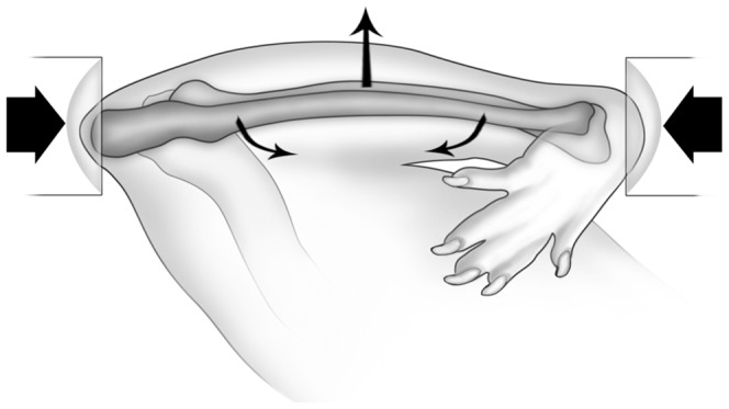
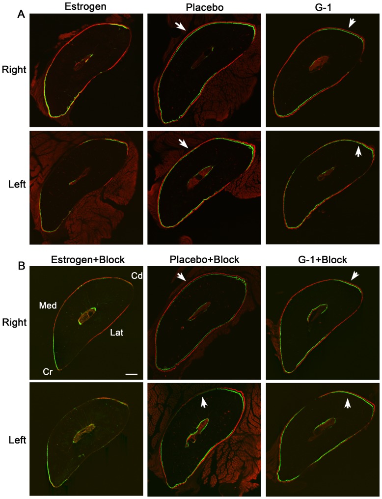
METHODOLOGY/PRINCIPAL FINDINGS: We used the rat ulna end-loading model to induce an adaptive modeling response in ovariectomized (OVX) female Sprague-Dawley rats. Rats were treated with a placebo, estrogen (17β-estradiol), or G-1, a GPER-specific agonist. Fourteen days after OVX, rats underwent unilateral cyclic loading of the right ulna; half of the rats in each group had brachial plexus anesthesia (BPA) of the loaded limb before loading. Ten days after loading, serum estrogen concentrations, dorsal root ganglion (DRG) gene expression of ERα, ERβ, GPER, CGRPα, TRPV1, TRPV4 and TRPA1, and load-induced skeletal responses were quantified. We hypothesized that estrogen and G-1 treatment would influence skeletal responses to cyclic loading through a neuronal mechanism. We found that estrogen suppresses periosteal bone formation in female rats. This physiological effect is not GPER-mediated. We also found that absolute mechanosensitivity in female rats was decreased, when compared with male rats. Blocking of adaptive bone formation by BPA in Placebo OVX females was reduced.
Estrogen acts to decrease periosteal bone formation in female rats in vivo. This effect is not GPER-mediated. Gender differences in absolute bone mechanosensitivity exist in young Sprague-Dawley rats with reduced mechanosensitivity in females, although underlying bone formation rate associated with growth likely influences this observation. In contrast to female and male rats, central neuronal signals had a diminished effect on adaptive bone formation in estrogen-deficient female rats.
性激素对骨骼有直接影响。雌激素通过经典的基因组雌激素受体α和β(ERα和 ERβ)、膜雌激素受体和非基因组 G 蛋白偶联雌激素受体(GPER)作用于骨骼。GPER 分布于整个神经系统,但对其对骨骼的影响知之甚少。在雄性大鼠中,对加载的适应是由神经元调节的,但在雌性大鼠中尚未对此进行研究。
方法/主要发现:我们使用大鼠尺骨末端加载模型诱导去卵巢(OVX)雌性 Sprague-Dawley 大鼠产生适应性建模反应。大鼠接受安慰剂、雌激素(17β-雌二醇)或 G-1(一种特定的 GPER 激动剂)治疗。OVX 后 14 天,大鼠右侧尺骨进行单侧循环加载;每组一半大鼠在加载前进行加载肢体臂丛神经阻滞(BPA)。加载后 10 天,测量血清雌激素浓度、背根神经节(DRG)中 ERα、ERβ、GPER、CGRPα、TRPV1、TRPV4 和 TRPA1 的基因表达,以及加载引起的骨骼反应。我们假设雌激素和 G-1 治疗会通过神经元机制影响骨骼对循环加载的反应。我们发现雌激素抑制雌性大鼠的骨膜骨形成。这种生理效应不是由 GPER 介导的。我们还发现,与雄性大鼠相比,雌性大鼠的绝对机械敏感性降低。在安慰剂 OVX 雌性大鼠中,BPA 阻断适应性骨形成的作用减弱。
雌激素在体内作用于减少雌性大鼠的骨膜骨形成。这种作用不是由 GPER 介导的。在年轻的 Sprague-Dawley 大鼠中,存在性别差异的绝对骨机械敏感性,雌性大鼠的机械敏感性降低,尽管与生长相关的基础骨形成率可能影响这一观察结果。与雌性和雄性大鼠不同,雌激素缺乏的雌性大鼠中,中枢神经元信号对适应性骨形成的影响减弱。