Tissue Engineering Laboratory and Berlin-Brandenburg Center for Regenerative Therapies, Department of Rheumatology and Clinical Immunology, Charité-Universitätsmedizin Berlin, Föhrer Strasse 15, Berlin 13353, Germany.
BMC Musculoskelet Disord. 2012 Sep 17;13:175. doi: 10.1186/1471-2474-13-175.
Tissue adhesives are useful means for various medical procedures. Since varying requirements cause that a single adhesive cannot meet all needs, bond strength testing remains one of the key applications used to screen for new products and study the influence of experimental variables. This study was conducted to develop an easy to use method to screen and evaluate tissue adhesives for tissue engineering applications.
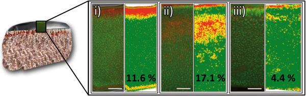
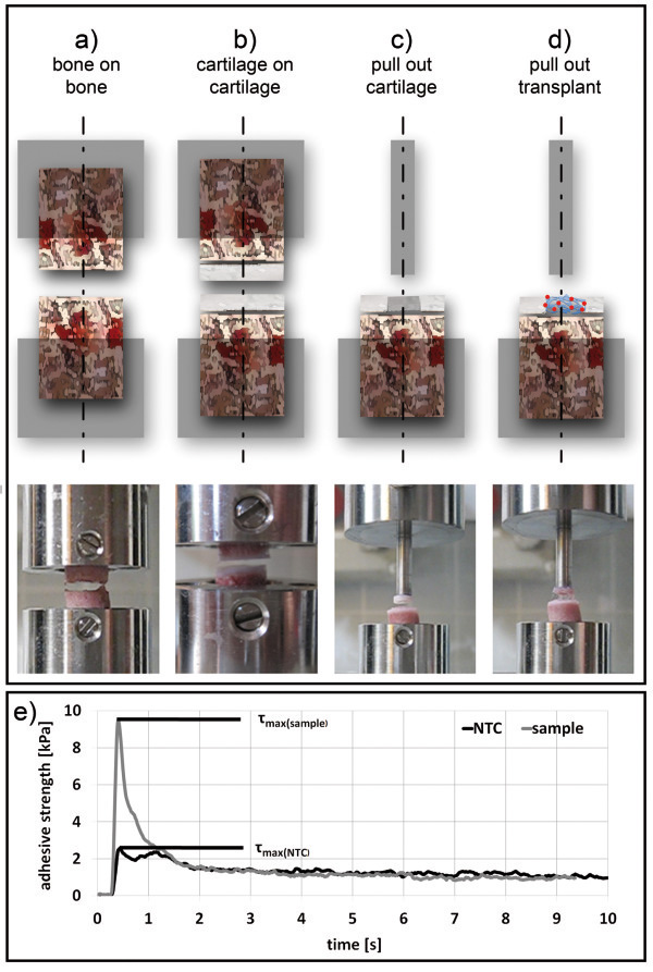
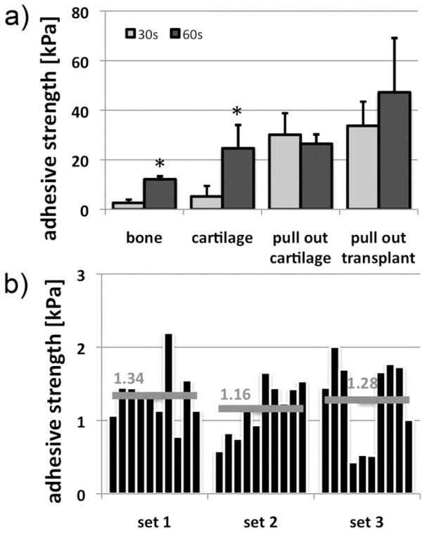
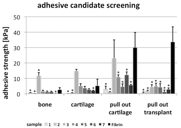
Tissue grips were designed to facilitate the reproducible production of substrate tissue and adhesive strength measurements in universal testing machines. Porcine femoral condyles were used to generate osteochondral test tissue cylinders (substrates) of different shapes. Viability of substrates was tested using PI/FDA staining. Self-bonding properties were determined to examine reusability of substrates (n = 3). Serial measurements (n = 5) in different operation modes (OM) were performed to analyze the bonding strength of tissue adhesives in bone (OM-1) and cartilage tissue either in isolation (OM-2) or under specific requirements in joint repair such as filling cartilage defects with clinical applied fibrin/PLGA-cell-transplants (OM-3) or tissues (OM-4). The efficiency of the method was determined on the basis of adhesive properties of fibrin glue for different assembly times (30 s, 60 s). Seven randomly generated collagen formulations were analyzed to examine the potential of method to identify new tissue adhesives.
Viability analysis of test tissue cylinders revealed vital cells (>80%) in cartilage components even 48 h post preparation. Reuse (n = 10) of test substrate did not significantly change adhesive characteristics. Adhesive strength of fibrin varied in different test settings (OM-1: 7.1 kPa, OM-2: 2.6 kPa, OM-3: 32.7 kPa, OM-4: 30.1 kPa) and was increasing with assembly time on average (2.4-fold). The screening of the different collagen formulations revealed a substance with significant higher adhesive strength on cartilage (14.8 kPa) and bone tissue (11.8 kPa) compared to fibrin and also considerable adhesive properties when filling defects with cartilage tissue (23.2 kPa).
The method confirmed adhesive properties of fibrin and demonstrated the dependence of adhesive properties and applied settings. Furthermore the method was suitable to screen for potential adhesives and to identify a promising candidate for cartilage and bone applications. The method can offer simple, replicable and efficient evaluation of adhesive properties in ex vivo specimens and may be a useful supplement to existing methods in clinical relevant settings.
组织粘合剂是各种医疗程序的有用手段。由于不同的需求导致单一的粘合剂无法满足所有需求,因此粘合强度测试仍然是筛选新产品和研究实验变量影响的关键应用之一。本研究旨在开发一种易于使用的方法,用于筛选和评估组织工程应用中的组织粘合剂。
设计了组织夹具,以方便在万能试验机中重复生产基底组织和粘合强度测量。使用猪股骨髁生成不同形状的骨软骨测试组织圆柱(基底)。使用 PI/FDA 染色测试基底的活力。确定自粘合特性以检查基底的可重复使用性(n=3)。进行了(n=5)不同操作模式(OM)的连续测量,以分析骨(OM-1)和软骨组织中组织粘合剂的粘合强度,要么单独分离(OM-2),要么在关节修复中特定要求下,如用临床应用的纤维蛋白/PLGA-细胞移植物(OM-3)或组织(OM-4)填充软骨缺陷。基于不同组装时间(30s,60s)下纤维蛋白胶的粘合性能,确定了该方法的效率。分析了七种随机生成的胶原配方,以检验该方法识别新的组织粘合剂的潜力。
测试组织圆柱的活力分析显示,即使在制备后 48 小时,软骨成分中的活细胞(>80%)。测试基底的重复使用(n=10)并没有显著改变粘合特性。纤维蛋白的粘合强度在不同的测试设置中有所不同(OM-1:7.1kPa,OM-2:2.6kPa,OM-3:32.7kPa,OM-4:30.1kPa),并且随着组装时间的增加而平均增加(2.4 倍)。不同胶原配方的筛选显示,一种物质在软骨(14.8kPa)和骨组织(11.8kPa)上的粘合强度明显高于纤维蛋白,并且在填充软骨组织缺陷时也具有相当的粘合性能(23.2kPa)。
该方法证实了纤维蛋白的粘合性能,并证明了粘合性能和应用设置的依赖性。此外,该方法适用于筛选潜在的粘合剂,并确定一种有前途的用于软骨和骨应用的候选物。该方法可提供简单、可重复和有效的体外标本粘合性能评估,可能是临床相关环境中现有方法的有用补充。