UCL Institute of Cognitive Neuroscience, 17 Queen Square, London, UK.
Neuroimage. 2013 Jan 1;64:722-7. doi: 10.1016/j.neuroimage.2012.08.076. Epub 2012 Sep 8.
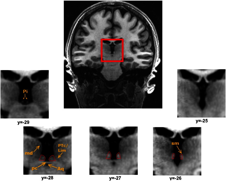
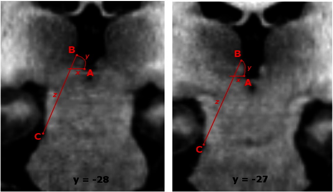
Recently there has been renewed interest in the habenula; a pair of small, highly evolutionarily conserved epithalamic nuclei adjacent to the medial dorsal (MD) nucleus of the thalamus. The habenula has been implicated in a range of behaviours including sleep, stress and pain, and studies in non-human primates have suggested a potentially important role in reinforcement processing, putatively via its effects on monoaminergic neurotransmission. Over the last decade, an increasing number of neuroimaging studies have reported functional responses in the human habenula using functional magnetic resonance imaging (fMRI). However, standard fMRI analysis approaches face several challenges in isolating signal from this structure because of its relatively small size, around 30 mm(3) in volume. In this paper we offer a set of guidelines for locating and manually tracing the habenula in humans using high-resolution T1-weighted structural images. We also offer recommendations for appropriate pre-processing and analysis of high-resolution functional magnetic resonance imaging (fMRI) data such that signal from the habenula can be accurately resolved from that in surrounding structures.
最近人们对缰核重新产生了兴趣;缰核是一对紧邻丘脑内侧背核(MD)的小型、高度进化保守的上丘脑核。缰核与包括睡眠、压力和疼痛在内的一系列行为有关,并且在非人类灵长类动物中的研究表明,它可能通过对单胺能神经传递的影响在强化处理中发挥重要作用。在过去的十年中,越来越多的使用功能磁共振成像(fMRI)的神经影像学研究报告了人类缰核的功能反应。然而,由于其相对较小的体积(约 30 立方毫米),标准 fMRI 分析方法在从该结构中分离信号时面临着一些挑战。在本文中,我们提供了一套使用高分辨率 T1 加权结构图像定位和手动追踪人类缰核的指南。我们还提供了有关高分辨率功能磁共振成像(fMRI)数据的适当预处理和分析的建议,以便能够准确地从周围结构中分辨出缰核的信号。