Kashani Mehdi, Farhadi Sareh, Rastegarfard Neda
Assistant Professor, Department of Orthodontics, Faculty of Dentistry, Shahed University, Tehran, Iran.
J Dent Res Dent Clin Dent Prospects. 2012 Summer;6(3):89-93. doi: 10.5681/joddd.2012.019. Epub 2012 Sep 1.
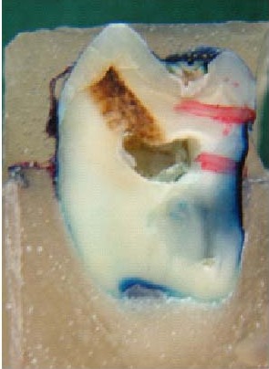
This in vitro study was designed to compare enamel demineralization depths adjacent to bands cemented with zinc polycarboxylate, glass ionomer (GI) and resin-modified glass ionomer (RMGI), in order to achieve minimal enamel demineralization during orthodontic treatment.
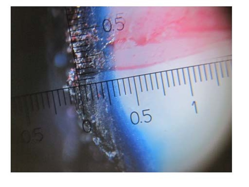
Sixty fully developed extracted third molars were randomly divided into three testgroups each containing 20 samples, used to cement orthodontic bands with zinc polycarboxylate, GI and RMGI. All samples were demineralized using White method using hydroxyapatite, latic acid and Carbapol for in vitro caries simulation, and then, immersed in 10% solution of methylene blue. The mean depth of dye penetration was assessed up to 0.1 millimeter, reflect-ing the depth of enamel demineralization. One way ANOVA and LSD statistical tests were employed to evaluate significant differences among groups.
The highest dye penetration depth was seen in zinc polycarboxylate group, followed by GI, and RMGI groups, respectively, with significant differences among each two groups (P < 0.05).
The use of RMGI cement seems to present significantly better prevention of enamel demineralization adja-cent to orthodontics bands.
本体外研究旨在比较用聚羧酸锌、玻璃离子水门汀(GI)和树脂改性玻璃离子水门汀(RMGI)粘结带环附近的牙釉质脱矿深度,以便在正畸治疗期间使牙釉质脱矿降至最低。
60颗完全发育的拔除第三磨牙随机分为三个试验组,每组20个样本,分别用于用聚羧酸锌、GI和RMGI粘结正畸带环。所有样本采用White方法,使用羟基磷灰石、乳酸和卡波姆进行体外龋齿模拟脱矿,然后浸入10%的亚甲蓝溶液中。评估染料渗透的平均深度至0.1毫米,反映牙釉质脱矿深度。采用单因素方差分析和LSD统计检验评估各组间的显著差异。
聚羧酸锌组的染料渗透深度最高,其次分别是GI组和RMGI组,两两之间差异显著(P < 0.05)。
使用RMGI粘结剂似乎能显著更好地预防正畸带环附近的牙釉质脱矿。