Molecular Oncology Group, Portuguese Institute of Oncology, Ed, Laboratórios-Piso 4, Rua Dr, António Bernardino de Almeida 4200-072, Porto, Portugal.
BMC Med. 2012 Sep 25;10:108. doi: 10.1186/1741-7015-10-108.
Periprostatic (PP) adipose tissue surrounds the prostate, an organ with a high predisposition to become malignant. Frequently, growing prostatic tumor cells extend beyond the prostatic organ towards this fat depot. This study aimed to determine the genome-wide expression of genes in PP adipose tissue in obesity/overweight (OB/OW) and prostate cancer patients.
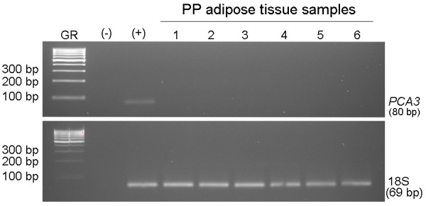
Differentially expressed genes in human PP adipose tissue were identified using microarrays. Analyses were conducted according to the donors' body mass index characteristics (OB/OW versus lean) and prostate disease (extra prostatic cancer versus organ confined prostate cancer versus benign prostatic hyperplasia). Selected genes with altered expression were validated by real-time PCR. Ingenuity Pathway Analysis (IPA) was used to investigate gene ontology, canonical pathways and functional networks.
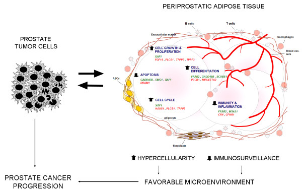
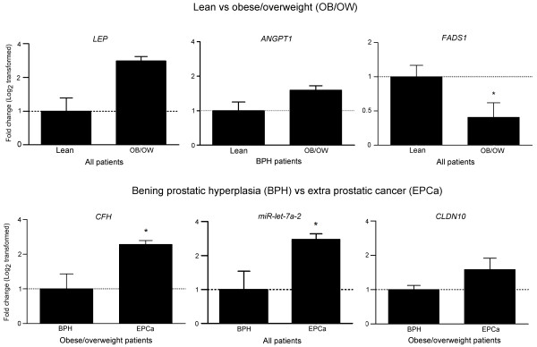
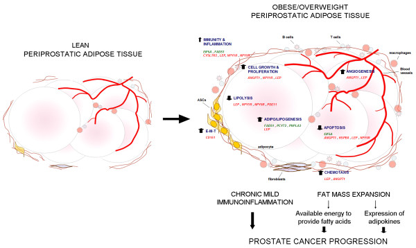
In the PP adipose tissue of OB/OW subjects, we found altered expression of genes encoding molecules involved in adipogenic/anti-lipolytic, proliferative/anti-apoptotic, and mild immunoinflammatory processes (for example, FADS1, down-regulated, and LEP and ANGPT1, both up-regulated). Conversely, in the PP adipose tissue of subjects with prostate cancer, altered genes were related to adipose tissue cellular activity (increased cell proliferation/differentiation, cell cycle activation and anti-apoptosis), whereas a downward impact on immunity and inflammation was also observed, mostly related to the complement (down-regulation of CFH). Interestingly, we found that the microRNA MIRLET7A2 was overexpressed in the PP adipose tissue of prostate cancer patients.
Obesity and excess adiposity modified the expression of PP adipose tissue genes to ultimately foster fat mass growth. In patients with prostate cancer the expression profile of PP adipose tissue accounted for hypercellularity and reduced immunosurveillance. Both findings may be liable to promote a favorable environment for prostate cancer progression.
前列腺周围脂肪组织(PP 脂肪组织)环绕着前列腺,前列腺极易发生恶性转化。生长中的前列腺肿瘤细胞常常会超出前列腺器官,延伸到这个脂肪库。本研究旨在确定肥胖/超重(OB/OW)和前列腺癌患者 PP 脂肪组织中的基因全基因组表达。
使用微阵列鉴定人 PP 脂肪组织中的差异表达基因。根据供体的体重指数特征(OB/OW 与瘦)和前列腺疾病(前列腺外癌与器官局限前列腺癌与良性前列腺增生)进行分析。通过实时 PCR 验证具有改变表达的选定基因。使用 ingenuity 通路分析(IPA)来研究基因本体论、经典途径和功能网络。
在 OB/OW 受试者的 PP 脂肪组织中,我们发现参与脂肪生成/抗脂肪分解、增殖/抗凋亡和轻度免疫炎症过程的基因表达发生改变(例如,下调 FADS1,上调 LEP 和 ANGPT1)。相反,在前列腺癌受试者的 PP 脂肪组织中,改变的基因与脂肪组织细胞活性有关(细胞增殖/分化增加、细胞周期激活和抗凋亡),同时也观察到对免疫和炎症的向下影响,主要与补体有关(下调 CFH)。有趣的是,我们发现微小 RNA MIRLET7A2 在前列腺癌患者的 PP 脂肪组织中过表达。
肥胖和多余的肥胖改变了 PP 脂肪组织基因的表达,最终促进了脂肪量的增长。在前列腺癌患者中,PP 脂肪组织的表达谱解释了细胞过度生长和免疫监视减少。这两种发现都可能容易促进前列腺癌进展的有利环境。