Department of Nuclear Medicine, University Hospital Muenster, Albert-Schweitzer-Campus 1, Gebäude A1, Muenster, 48149, Germany.
EJNMMI Res. 2012 Sep 27;2(1):51. doi: 10.1186/2191-219X-2-51.
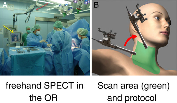
Freehand single photon emission computed tomography (fSPECT) is a three-dimensional (3-D) tomographic imaging modality based on data acquisition with a handheld detector that is moved freely, in contrast to conventional, gantry-mounted gamma camera systems. In this pilot study, we evaluated the feasibility of fSPECT for intraoperative 3-D mapping in patients with parathyroid adenomas.
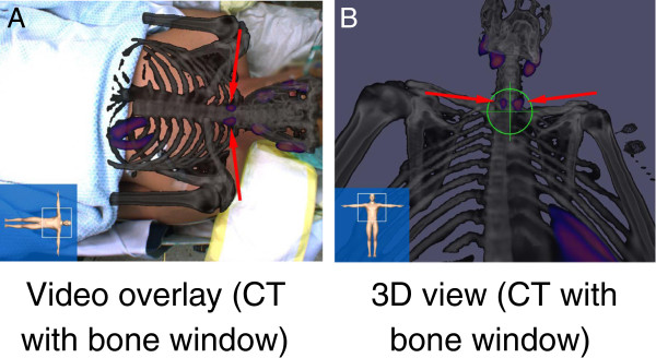
Three patients (range 30 to 45 years) diagnosed with hyperparathyroidism (one primary and two tertiary) underwent parathyroid scintigraphy with technetium-99m sestamibi (99mTc-MIBI) to localize parathyroid adenomas. Two patients were referred with persistent hyperparathyroidism after conventional parathyroidectomy. In all three patients, a planar scintigraphy of the neck was performed 10 min after injection (p.i.) followed by SPECT/CT (Symbia T2, Siemens Healthcare) and a correlative ultrasound 2 h p.i. 99mTc-MIBI scan was performed the day before surgery in two patients and at the same day in one patient. fSPECT images were acquired intraoperatively using declipse SPECT (SurgicEyeTM).
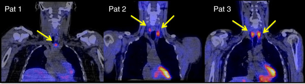
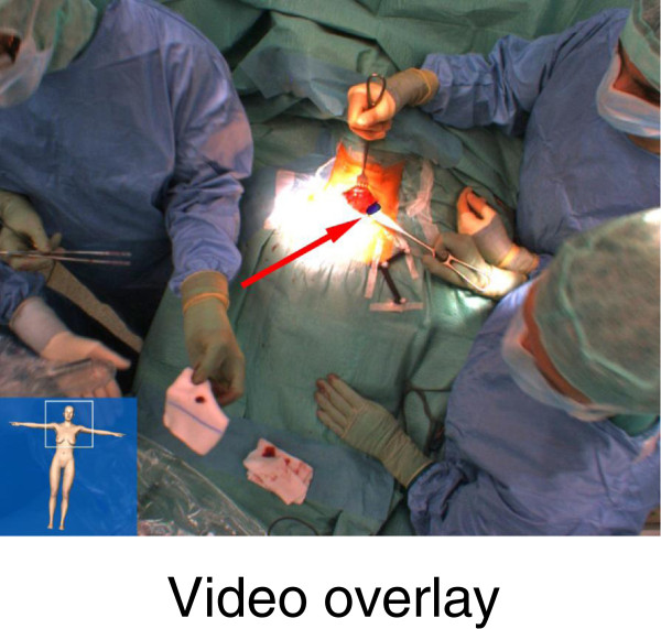
A total of five parathyroid adenomas were successfully located with SPECT/CT. fSPECT allowed intraoperative detection of all adenomas, and successful parathyroidectomy was accomplished. Parathyroid hormone level decreased intraoperatively in all three patients, on average, by 79% (range 72% to 91%).
In this preliminary study, we could demonstrate that intraoperative localization of parathyroid adenomas is feasible using the freehand SPECT technology, thus allowing an image-guided parathyroidectomy.
自由手单光子发射计算机断层扫描(fSPECT)是一种基于手持探测器自由移动的数据采集的三维(3-D)断层成像方式,与传统的、安装在龙门架上的伽马相机系统不同。在这项初步研究中,我们评估了 fSPECT 用于甲状旁腺腺瘤患者术中 3-D 定位的可行性。
三名(年龄 30 至 45 岁)被诊断为甲状旁腺功能亢进症的患者(原发性和三发性各一名)接受了锝-99m 甲氧基异丁基异腈(99mTc-MIBI)甲状旁腺闪烁扫描以定位甲状旁腺腺瘤。两名患者在常规甲状旁腺切除术后因持续性甲状旁腺功能亢进而被转诊。在所有三名患者中,在注射后 10 分钟(p.i.)进行颈平面闪烁扫描,随后进行 SPECT/CT(Siemens Healthcare 的 Symbia T2)和 2 小时 p.i.的相关超声检查。在两名患者中,99mTc-MIBI 扫描在手术前一天进行,在一名患者中在同一天进行。在术中使用 declipse SPECT(SurgicEyeTM)获取 fSPECT 图像。
SPECT/CT 成功定位了总共五个甲状旁腺腺瘤。fSPECT 允许术中检测到所有腺瘤,并成功进行了甲状旁腺切除术。在所有三名患者中,甲状旁腺激素水平在术中平均降低了 79%(范围为 72%至 91%)。
在这项初步研究中,我们能够证明使用自由手 SPECT 技术进行术中甲状旁腺腺瘤定位是可行的,从而实现了图像引导的甲状旁腺切除术。