Department of Cerebral Ischemia and Neurodegeneration, Institut d'Investigacions Biomèdiques de Barcelona-Consejo Superior de Investigaciones Científicas (CSIC), Institut d'Investigacions Biomèdiques August-Pi i Sunyer (IDIBAPS), Barcelona, Spain.
PLoS One. 2012;7(9):e45227. doi: 10.1371/journal.pone.0045227. Epub 2012 Sep 20.
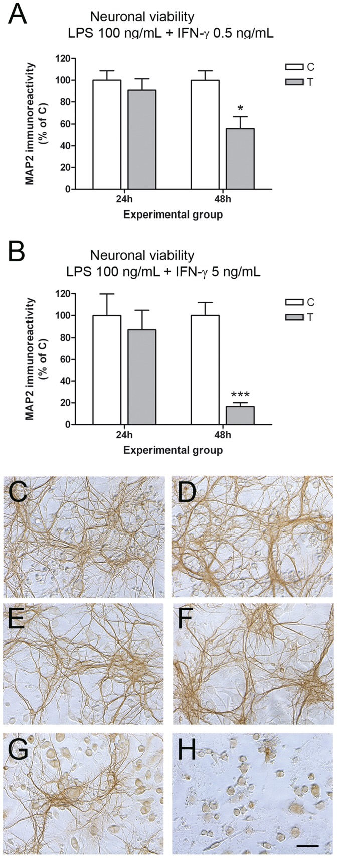
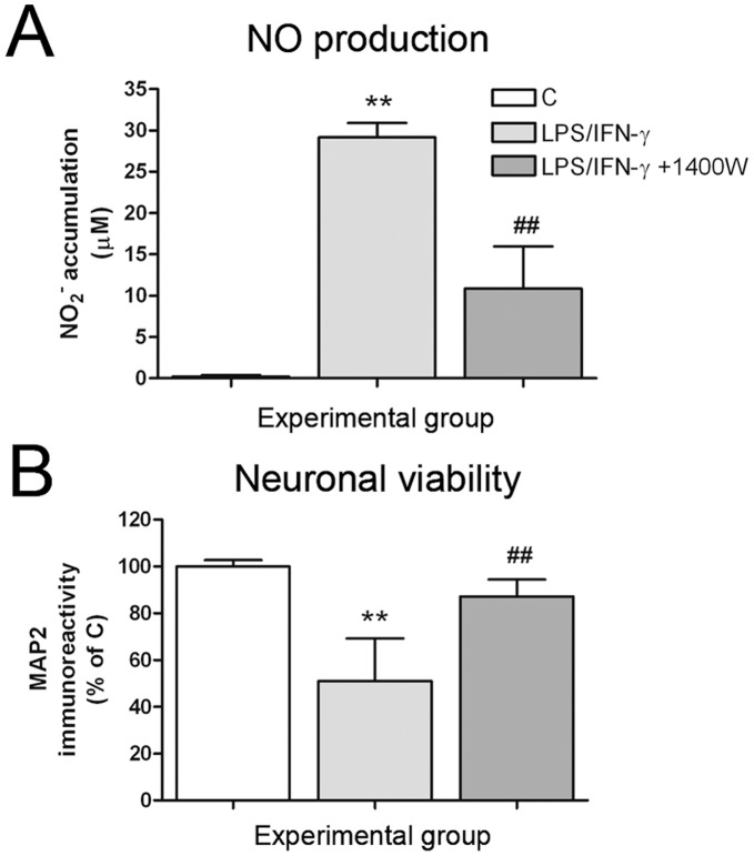
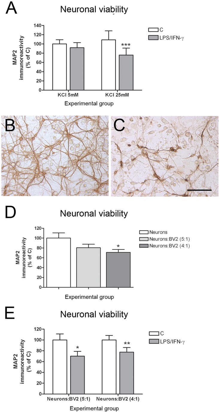
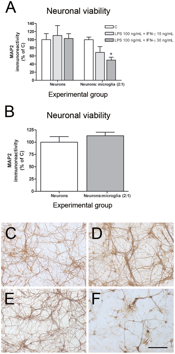
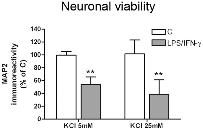
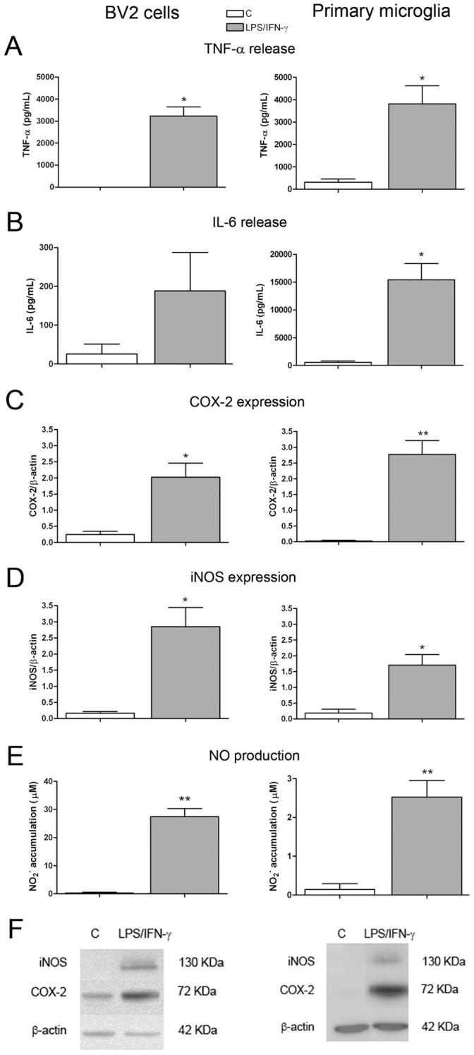
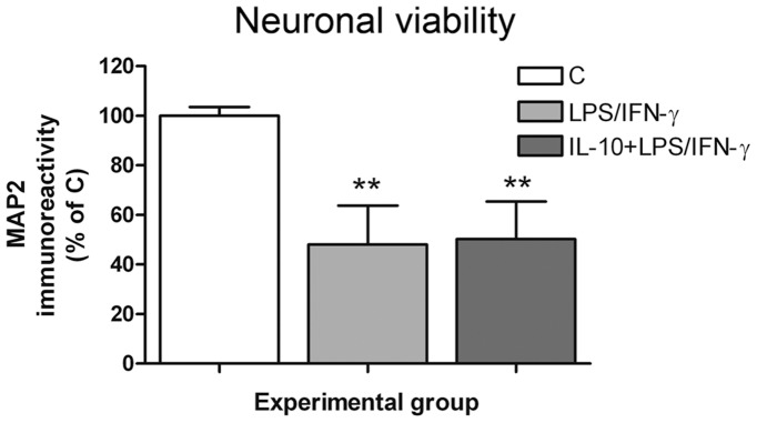
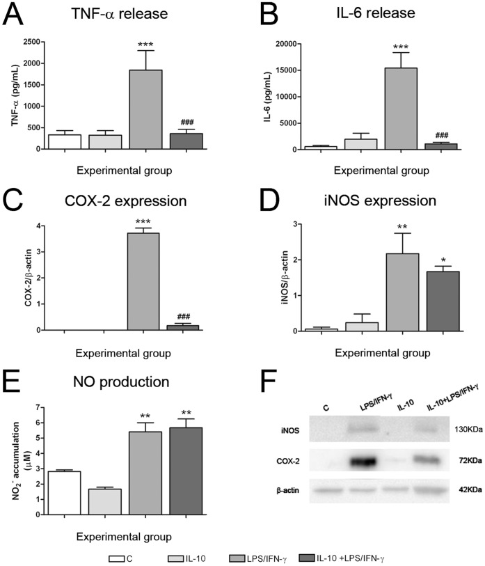
Neuron-microglia co-cultures treated with pro-inflammatory agents are a useful tool to study neuroinflammation in vitro, where to test the potential neuroprotective effect of anti-inflammatory compounds. However, a great diversity of experimental conditions can be found in the literature, making difficult to select the working conditions when considering this approach for the first time. We compared the use of neuron-primary microglia and neuron-BV2 cells (a microglial cell line) co-cultures, using different neuron:microglia ratios, treatments and time post-treatment to induce glial activation and derived neurotoxicity. We show that each model requires different experimental conditions, but that both neuron-BV2 and neuron-primary microglia LPS/IFN-γ-treated co-cultures are good to study the potential neuroprotective effect of anti-inflammatory agents. The contribution of different pro-inflammatory parameters in the neurotoxicity induced by reactive microglial cells was determined. IL-10 pre-treatment completely inhibited LPS/IFN-γ-induced TNF-α and IL-6 release, and COX-2 expression both in BV2 and primary microglial cultures, but not NO production and iNOS expression. However, LPS/IFN-γ induced neurotoxicity was not inhibited in IL-10 pre-treated co-cultures. The inhibition of NO production using the specific iNOS inhibitor 1400 W totally abolished the neurotoxic effect of LPS/IFN-γ, suggesting a major role for NO in the neurotoxic effect of activated microglia. Consequently, among the anti-inflammatory agents, special attention should be paid to compounds that inhibit NO production.
用促炎剂处理的神经元-小胶质细胞共培养物是研究体外神经炎症的有用工具,可用于测试抗炎化合物的潜在神经保护作用。然而,文献中存在着极大的实验条件多样性,使得在首次考虑这种方法时难以选择工作条件。我们比较了神经元-原代小胶质细胞和神经元-BV2 细胞(小胶质细胞系)共培养物的使用,使用不同的神经元:小胶质细胞比例、处理和处理后时间来诱导小胶质细胞激活和衍生的神经毒性。我们表明,每种模型都需要不同的实验条件,但神经元-BV2 和神经元-原代小胶质细胞 LPS/IFN-γ 处理的共培养物都适合研究抗炎剂的潜在神经保护作用。确定了不同促炎参数在反应性小胶质细胞诱导的神经毒性中的作用。IL-10 预处理完全抑制了 LPS/IFN-γ 诱导的 TNF-α 和 IL-6 释放以及 BV2 和原代小胶质细胞培养物中的 COX-2 表达,但不抑制 NO 产生和 iNOS 表达。然而,在 IL-10 预处理的共培养物中,LPS/IFN-γ 诱导的神经毒性并未被抑制。使用特异性 iNOS 抑制剂 1400 W 抑制 NO 产生完全消除了 LPS/IFN-γ 的神经毒性作用,表明 NO 在激活的小胶质细胞的神经毒性作用中起主要作用。因此,在抗炎剂中,应特别注意抑制 NO 产生的化合物。