Prabhasawat Pinnita, Ekpo Pattama, Uiprasertkul Mongkol, Chotikavanich Suksri, Tesavibul Nattaporn
Department of Ophthalmology, Faculty of Medicine, Siriraj Hospital, Mahidol University, Bangkok, Thailand.
Clin Ophthalmol. 2012;6:1483-92. doi: 10.2147/OPTH.S33951. Epub 2012 Sep 11.
To investigate the clinical outcomes of cultivated corneal limbal epithelial transplantation (CLET) using human amniotic membrane for corneal limbal stem-cell deficiency.
Prospective, noncomparative case series. Eighteen patients (19 eyes) with severe ocular surface diseases were chosen to undergo CLET using human amniotic membrane. Twelve eyes received auto-CLET, and seven eyes received allo-CLET. Clinical outcomes of corneal surface epithelialization, conjunctivalization, inflammation, visual acuity, graft status, and complications were observed.
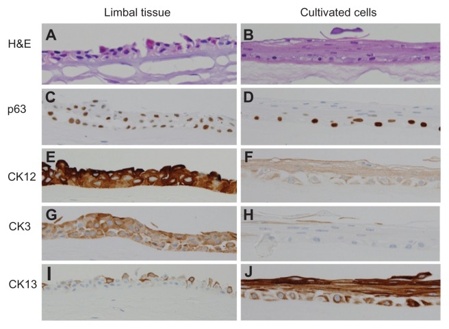
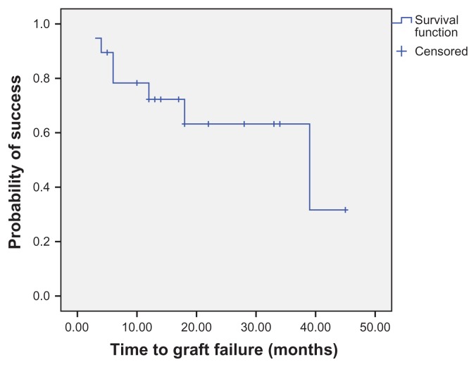
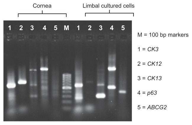
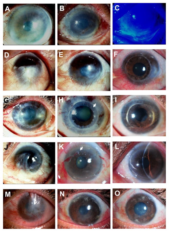
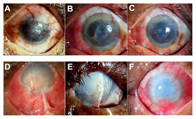
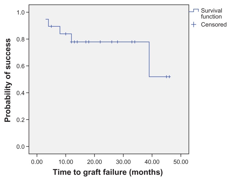
Corneal epithelium cultivated on amniotic membrane (two to four layers) was positive for molecular markers p63, ABCG2, CK3, and CK12. The mean patient age was 44.7 ± 15.2 years. A successful clinical outcome, defined as corneal epithelialization without central conjunctivalization or severe inflammation, was obtained in 14 (73.7%) of 19 eyes (mean follow-up 26.1 ± 13.5 months; range 6-47). A histopathologic success, defined as absence of goblet cells at the central cornea, was achieved in 12 (63.2%) eyes. Clinical failures occurred in five (26.3%) of 19 eyes, and histopathologic failures occurred in seven (36.8%) of 19 eyes. Survival analysis at 1 year showed that the clinical success rate was 77.9% and the pathological success rate was 72.3%. Fourteen of 19 (73.7%) eyes had visual acuity improvements after CLET. Six cases underwent penetrating keratoplasty; five of these grafts remained clear after 20.4 ± 6.9 months (range, 12-31) of follow-up. Complications included infectious keratitis (three cases) and recurrent symblepharon (one case). All complicated cases had lid abnormalities. Factors affecting the final clinical outcomes were lid abnormalities, abnormal corneal stromal beds, and complications.
CLET can successfully restore ocular surface damage in most cases with corneal limbal stem cell deficiency.
探讨使用人羊膜的培养角膜缘上皮移植术(CLET)治疗角膜缘干细胞缺乏症的临床疗效。
前瞻性、非对照病例系列研究。选取18例(19眼)患有严重眼表疾病的患者接受使用人羊膜的CLET治疗。12眼接受自体CLET,7眼接受异体CLET。观察角膜表面上皮化、结膜化、炎症、视力、植片情况及并发症等临床疗效。
在羊膜上培养的角膜上皮(2至4层)分子标志物p63、ABCG2、CK3和CK12呈阳性。患者平均年龄为44.7±15.2岁。19眼中有14眼(73.7%)获得了成功的临床结局,定义为角膜上皮化且无中央结膜化或严重炎症(平均随访26.1±13.5个月;范围6 - 47个月)。12眼(63.2%)实现了组织病理学成功,定义为中央角膜无杯状细胞。19眼中有5眼(26.3%)出现临床失败,19眼中有7眼(36.8%)出现组织病理学失败。1年生存率分析显示临床成功率为77.9%,病理成功率为72.3%。19眼中有14眼(73.7%)在CLET后视力提高。6例接受了穿透性角膜移植术;其中5例移植片在随访20.4±6.9个月(范围12 - 31个月)后保持透明。并发症包括感染性角膜炎(3例)和复发性睑球粘连(1例)。所有复杂病例均有眼睑异常。影响最终临床结局的因素包括眼睑异常、角膜基质床异常和并发症。
CLET在大多数角膜缘干细胞缺乏症病例中可成功修复眼表损伤。