Department of Urology, Indiana University School of Medicine, Indianapolis, Indiana, United States of America.
PLoS One. 2012;7(10):e47417. doi: 10.1371/journal.pone.0047417. Epub 2012 Oct 12.
Interleukin 18 (IL-18) is a pro-inflammatory cytokine that mediates fibrotic renal injury during obstruction. Macrophages are a well-known source of IL-18; however, renal tubular epithelial cells are also a potential source of this cytokine. We hypothesized that IL-18 is predominantly a renal tubular cell product and is produced during renal obstruction independent of macrophage infiltration.
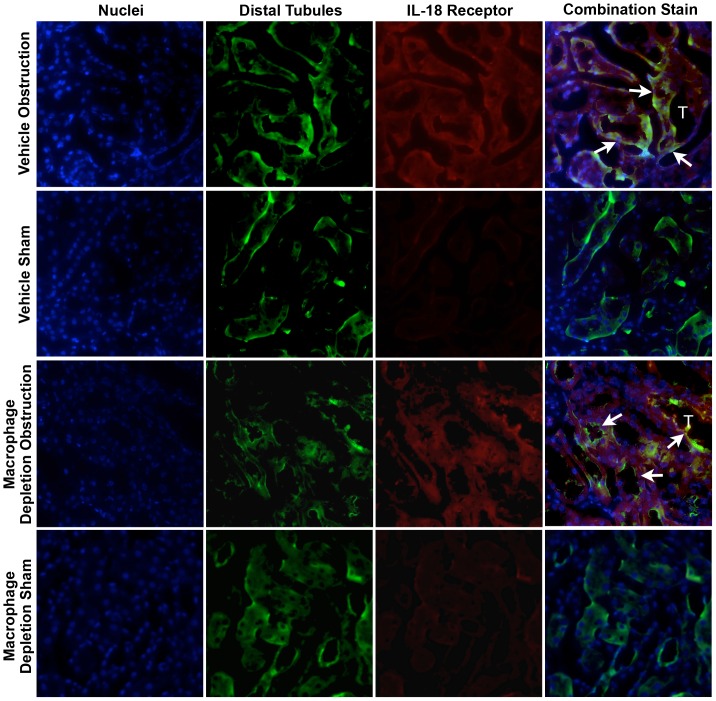
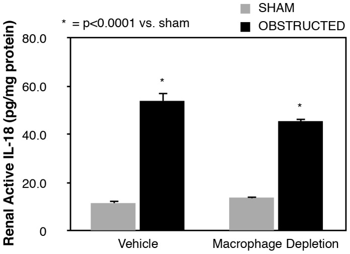
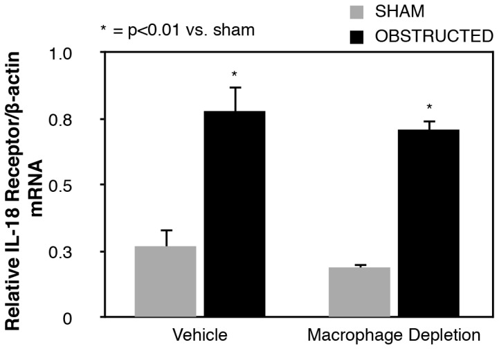
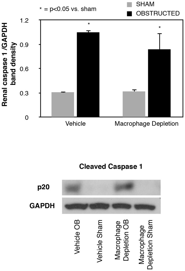
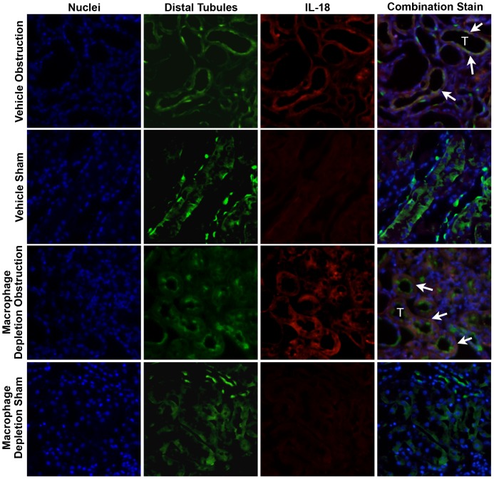
To study this, male C57BL6 mice were subjected to unilateral ureteral obstruction (UUO) vs. sham operation in the presence or absence of macrophage depletion (liposomal clodronate (1 ml/100 g body weight i.v.)). The animals were sacrificed 1 week after surgery and renal cortical tissue harvested. Tissue levels of active IL-18 (ELISA), IL-18 receptor mRNA expression (real time PCR), and active caspase-1 expression (western blot) were measured. The cellular localization of IL-18 and IL-18R was assessed using dual labeling immunofluorescent staining (IFS).
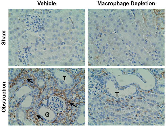
Immunohistochemical staining of renal tissue sections confirmed macrophage depletion by liposomal clodronate. IL-18 production, IL-18R expression, and active caspase 1 expression were elevated in response to renal obstruction and did not decline to a significant degree in the presence of macrophage depletion. Obstruction-induced IL-18 and IL-18R production localized predominantly to tubular epithelial cells (TEC) during obstruction despite macrophage depletion.
These results demonstrate that renal tubular epithelial cells are the primary source of IL-18 production during obstructive injury, and that tubular cell production of IL-18 occurs independent of macrophage infiltration.
白细胞介素 18(IL-18)是一种促炎细胞因子,可介导梗阻时的纤维化肾损伤。巨噬细胞是 IL-18 的主要来源;然而,肾小管上皮细胞也是这种细胞因子的潜在来源。我们假设 IL-18 主要是肾小管细胞的产物,并且在不依赖巨噬细胞浸润的情况下在肾梗阻期间产生。
为了研究这一点,雄性 C57BL6 小鼠接受单侧输尿管梗阻(UUO)与假手术对照,同时存在或不存在巨噬细胞耗竭(脂质体氯膦酸盐(1ml/100g 体重静脉内))。手术后 1 周处死动物并采集肾皮质组织。测量组织中活性 IL-18(ELISA)、IL-18 受体 mRNA 表达(实时 PCR)和活性半胱天冬酶-1 表达(western blot)。使用双标记免疫荧光染色(IFS)评估 IL-18 和 IL-18R 的细胞定位。
肾组织切片的免疫组织化学染色证实了脂质体氯膦酸盐对巨噬细胞的耗竭。IL-18 产生、IL-18R 表达和活性 caspase 1 表达在肾梗阻时升高,并且在巨噬细胞耗竭时并未显著降低。尽管存在巨噬细胞耗竭,但梗阻诱导的 IL-18 和 IL-18R 产生在梗阻期间主要定位于肾小管上皮细胞(TEC)。
这些结果表明,在梗阻性损伤期间,肾小管上皮细胞是 IL-18 产生的主要来源,并且肾小管细胞产生 IL-18 发生在巨噬细胞浸润之前。