Division of Gastroenterology, University of Pennsylvania Perelman School of Medicine, Philadelphia, Pennsylvania; Department of Medicine, University of Pennsylvania Perelman School of Medicine, Philadelphia, Pennsylvania; Abramson Cancer Center, University of Pennsylvania, Philadelphia, Pennsylvania.
Division of Nuclear Medicine and Molecular Imaging, Department of Radiology, Massachusetts General Hospital, Boston, Massachusetts.
Gastroenterology. 2013 Feb;144(2):294-297. doi: 10.1053/j.gastro.2012.10.030. Epub 2012 Oct 22.
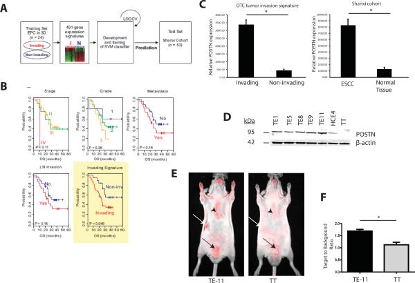
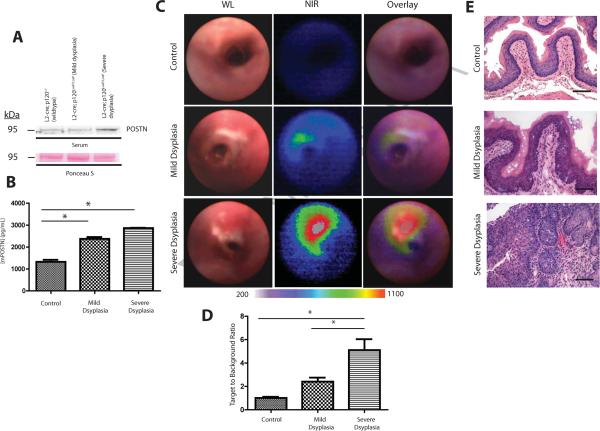
Imaging strategies that detect early stage esophageal squamous cell carcinoma (ESCC) could improve clinical outcomes, when combined with endoscopic approaches. Periostin is an integrin-binding protein that is important in the tumor microenvironment. We created a fluorescent-labeled antibody that recognizes periostin and binds specifically to ESCC xenograft tumors in mice. In L2-cre;p120ctnLoxP/LoxP mice, which develop squamous cell cancers that resemble human ESCC, we visualized the probe in preneoplastic and neoplastic esophageal lesions using near-infrared fluorescent imaging with upper-gastrointestinal endoscopy. Periostin might be a biomarker of the esophageal tumor microenvironment that can be used to detect preneoplastic lesions.
成像策略可以检测早期食管鳞状细胞癌(ESCC),与内镜方法结合使用时,可以改善临床结果。骨膜蛋白是一种整合素结合蛋白,在肿瘤微环境中具有重要作用。我们创建了一种荧光标记的抗体,可识别骨膜蛋白并特异性结合小鼠的 ESCC 异种移植肿瘤。在 L2-cre;p120ctnLoxP/LoxP 小鼠中,会发展出类似于人类 ESCC 的鳞状细胞癌,我们使用上消化道内镜的近红外荧光成像来可视化前病变和肿瘤性食管病变中的探针。骨膜蛋白可能是一种可以用于检测前病变的食管肿瘤微环境的生物标志物。