1. Theragnostic Laboratory, Department of Imaging & Pathology, Biomedical Sciences Group, KU Leuven, Herestraat 49, Leuven, Belgium. ; 2. Molecular Small Animal Imaging Centre/MoSAIC, Biomedical Sciences Group, KU Leuven, Herestraat 49, Leuven, Belgium.
Theranostics. 2012;2(10):1010-9. doi: 10.7150/thno.4924. Epub 2012 Oct 18.
The present animal experiments were conducted to evaluate radioiodinated Hypericin (Hyp) for its regional distribution as well as theranostic potentials.
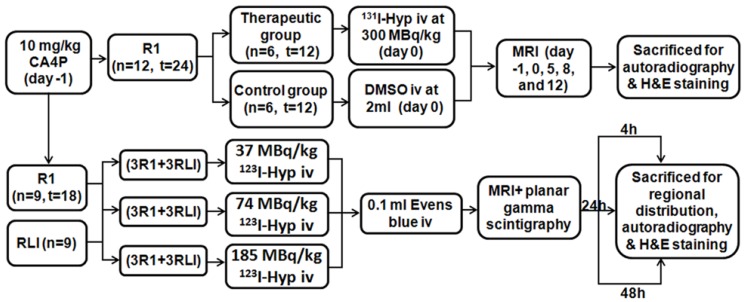
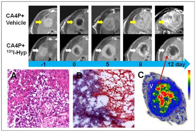
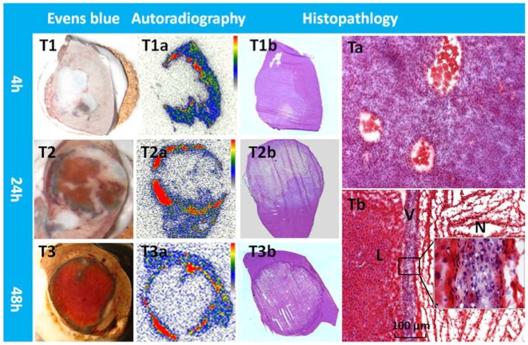
Rat models of reperfused liver infarction (RLI) and hepatic rhabdomyosarcoma (R1) were surgically induced. R1 models received Combretastatin A4 phosphate (CA4P) intravenously at 10 mg/kg 24 h prior to radioiodinated Hyp. Three groups of 6 rats each containing 3 RLI and 3 R1 models received iv injections of (123)I-Hyp at 37, 74, and 185 MBq/kg respectively and followed by 0.1 ml of 1% Evans blue solution were sacrificed at 4, 24 and 48 hour post injection immediately after in vivo examination of MRI and planar gamma scintigraphy. Besides, two groups of 6 R1 models that received either 300 MBq/kg of (131)I-Hyp or vehicle intravenously were examined using MRI to compare tumor growth for 12 days. Autoradiography, gamma counting, and histopathology were performed for postmortem verifications and quantification.
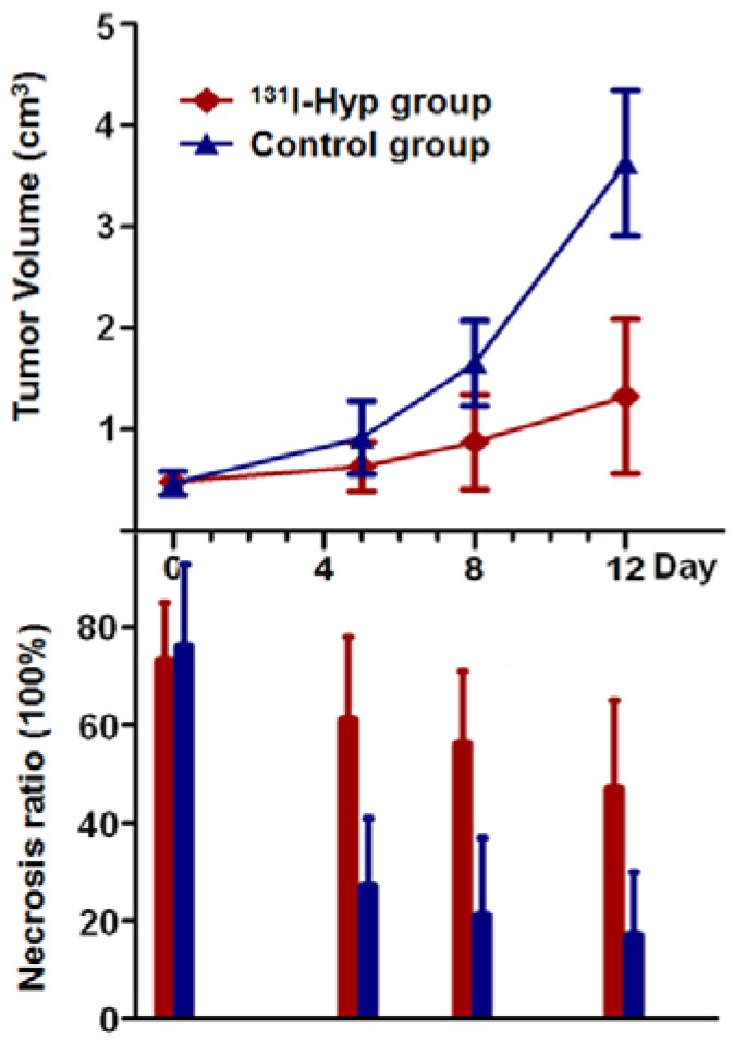
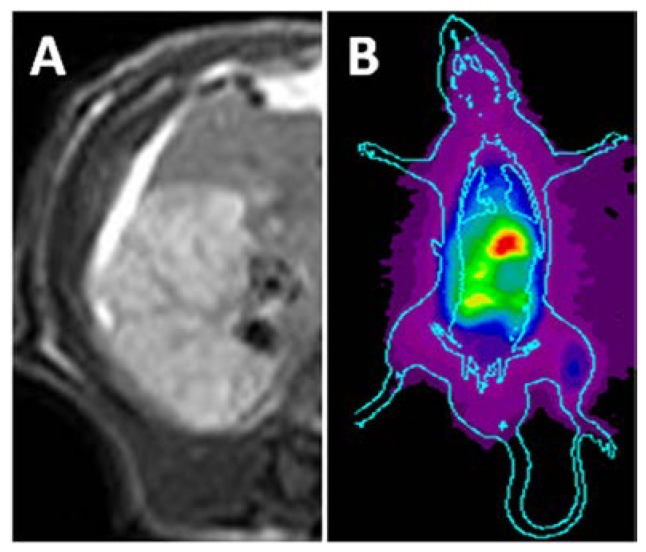
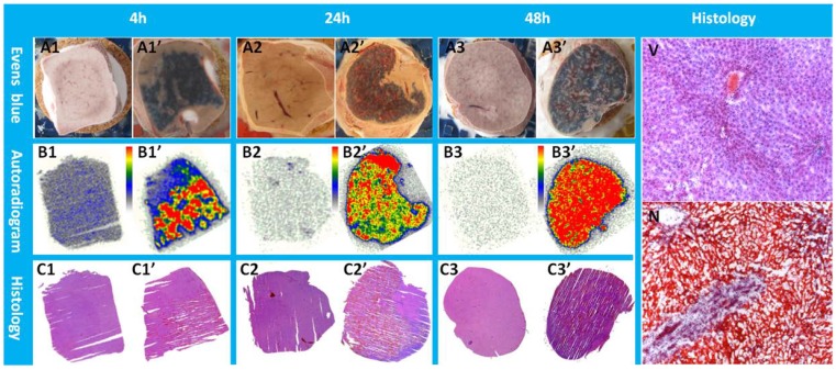
Necrosis as seen in vivo on contrast-enhanced MRI corresponded well with the hot spots on planar scintigraphy. Autoradiography and gamma counting revealed intense accumulation of (123)I-Hyp in necrotic liver (3.94 ± 1.60, 5.38 ± 1.04, and 6.03 ± 2.09 %ID/g ± SD) and necrotic tumor (4.27 ± 0.76, 5.57 ± 0.76, and 5.68 ± 1.33 %ID/g ± SD) relative to normal liver (1.76 ± 0.54, 0.41 ± 0.18, and 0.16 ± 0.07 %ID/g ± SD), with a high necrosis-to-liver ratio of 2.3, 14.0, and 37.0 at 4, 24 and 48 h respectively. Tumor volumes in R1 models that received (131)I-Hyp and vehicle changed from 0.45 ± 0.09, and 0.47 ± 0.12 cm(3) (p > 0.05) on day 0 to1.32 ± 0.76 and 3.63 ± 0.72 cm(3 )(p < 0.001) on day 12, with the corresponding necrosis ratios from 73 ± 12 %, and 76 ± 17 % to 47 ± 18% and 17 ± 13 % (p < 0.01), and with the tumor DT of 7.3 ± 1.0 and 4.2 ± 0.7 days, respectively.
Radioiodinated Hyp as a necrosis avid tracer appears promising for non-invasive imaging diagnosis of necrosis-related pathologies. Its prominent targetability to necrosis allows targeted radiotherapy for malignancies on top of a prior necrosis-inducing treatment.
本动物实验旨在评估放射性碘标记金丝桃素(Hyp)在肝再灌注梗死(RLI)和肝横纹肌肉瘤(R1)中的区域分布及其治疗潜力。
通过手术诱导 RLI 和 R1 大鼠模型。在接受放射性碘标记 Hyp 前 24 小时,R1 模型静脉注射 CA4P10mg/kg。每组 6 只大鼠,每组包含 3 只 RLI 和 3 只 R1 模型,分别静脉注射(123)I-Hyp37、74 和 185MBq/kg,然后立即在体内检查 MRI 和平面伽马闪烁成像后,于注射后 4、24 和 48 小时处死,各注射 0.1ml1% Evans 蓝溶液。此外,两组 6 只 R1 模型分别接受 300MBq/kg(131)I-Hyp 或静脉内载体,使用 MRI 检查 12 天以比较肿瘤生长情况。进行放射自显影、伽马计数和组织病理学检查以进行死后验证和定量。
MRI 上观察到的坏死与平面闪烁成像上的热点非常吻合。放射自显影和伽马计数显示,(123)I-Hyp 在坏死的肝脏(3.94±1.60、5.38±1.04 和 6.03±2.09%ID/g±SD)和坏死的肿瘤(4.27±0.76、5.57±0.76 和 5.68±1.33%ID/g±SD)中的积累非常强烈,与正常肝脏(1.76±0.54、0.41±0.18 和 0.16±0.07%ID/g±SD)相比,坏死与肝脏的比值分别为 2.3、14.0 和 37.0,在 4、24 和 48 小时分别为 2.3、14.0 和 37.0。接受(131)I-Hyp 和载体的 R1 模型的肿瘤体积从 0.45±0.09 和 0.47±0.12cm3(p>0.05)变为第 12 天的 1.32±0.76 和 3.63±0.72cm3(p<0.001),相应的坏死率从 73±12%和 76±17%变为 47±18%和 17±13%(p<0.01),肿瘤 DT 分别为 7.3±1.0 和 4.2±0.7 天。
放射性碘标记金丝桃素作为一种坏死亲和力示踪剂,在非侵入性成像诊断与坏死相关的病理方面似乎很有前景。它对坏死的显著靶向性允许在先前诱导坏死的治疗之上进行针对恶性肿瘤的靶向放射治疗。