Taylor Julia A, Richter Catherine A, Suzuki Atsuko, Watanabe Hajime, Iguchi Taisen, Coser Kathryn R, Shioda Toshihiro, vom Saal Frederick S
Division of Biological Sciences, University of Missouri, Columbia, Missouri, United States of America.
PLoS One. 2012;7(10):e48311. doi: 10.1371/journal.pone.0048311. Epub 2012 Oct 29.
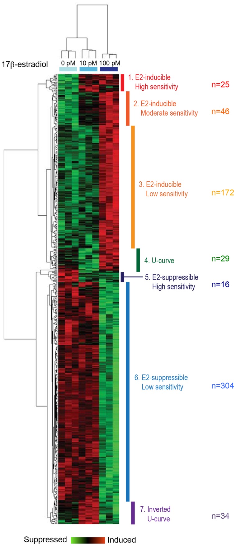
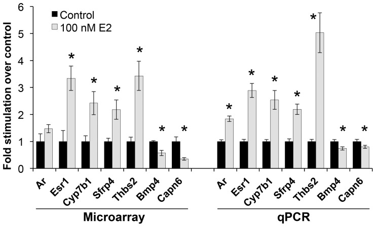
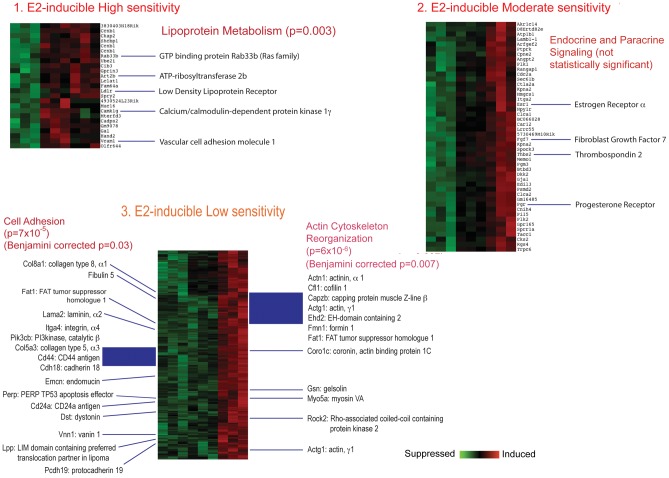
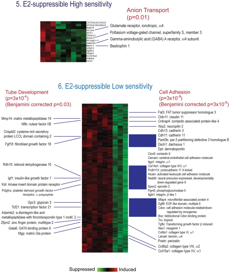
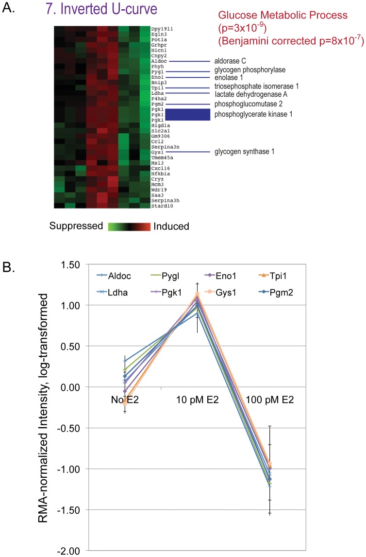
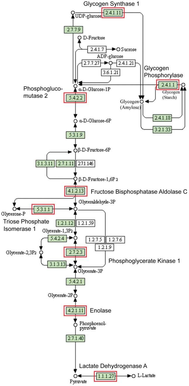
Developmental exposure of mouse fetuses to estrogens results in dose-dependent permanent effects on prostate morphology and function. Fetal prostatic mesenchyme cells express estrogen receptor alpha (ERα) and androgen receptors and convert stimuli from circulating estrogens and androgens into paracrine signaling to regulate epithelial cell proliferation and differentiation. To obtain mechanistic insight into the role of different doses of estradiol (E2) in regulating mesenchymal cells, we examined E2-induced transcriptomal changes in primary cultures of fetal mouse prostate mesenchymal cells. Urogenital sinus mesenchyme cells were obtained from male mouse fetuses at gestation day 17 and exposed to 10 pM, 100 pM or 100 nM E2 in the presence of a physiological concentration of dihydrotestosterone (0.69 nM) for four days. Gene ontology studies suggested that low doses of E2 (10 pM and 100 pM) induce genes involved in morphological tissue development and sterol biosynthesis but suppress genes involved in growth factor signaling. Genes involved in cell adhesion were enriched among both up-regulated and down-regulated genes. Genes showing inverted-U-shape dose responses (enhanced by E2 at 10 pM E2 but suppressed at 100 pM) were enriched in the glycolytic pathway. At the highest dose (100 nM), E2 induced genes enriched for cell adhesion, steroid hormone signaling and metabolism, cytokines and their receptors, cell-to-cell communication, Wnt signaling, and TGF- β signaling. These results suggest that prostate mesenchymal cells may regulate epithelial cells through direct cell contacts when estrogen level is low whereas secreted growth factors and cytokines might play significant roles when estrogen level is high.
小鼠胎儿在发育过程中暴露于雌激素会对前列腺形态和功能产生剂量依赖性的永久性影响。胎儿前列腺间充质细胞表达雌激素受体α(ERα)和雄激素受体,并将循环中的雌激素和雄激素刺激转化为旁分泌信号,以调节上皮细胞的增殖和分化。为了深入了解不同剂量的雌二醇(E2)在调节间充质细胞中的作用机制,我们检测了E2诱导的小鼠胎儿前列腺间充质细胞原代培养物中的转录组变化。在妊娠第17天从雄性小鼠胎儿中获取泌尿生殖窦间充质细胞,并在生理浓度的二氢睾酮(0.69 nM)存在下,将其暴露于10 pM、100 pM或100 nM的E2中4天。基因本体研究表明,低剂量的E2(10 pM和100 pM)诱导参与形态组织发育和甾醇生物合成的基因,但抑制参与生长因子信号传导的基因。参与细胞粘附的基因在上调基因和下调基因中均富集。显示倒U形剂量反应的基因(在10 pM E2时被E2增强,但在100 pM时被抑制)在糖酵解途径中富集。在最高剂量(100 nM)时,E2诱导的基因富集于细胞粘附、类固醇激素信号传导和代谢、细胞因子及其受体、细胞间通讯、Wnt信号传导和TGF-β信号传导。这些结果表明,当雌激素水平较低时,前列腺间充质细胞可能通过直接细胞接触来调节上皮细胞,而当雌激素水平较高时,分泌的生长因子和细胞因子可能发挥重要作用。