Han Jingquan, Cao Shouqiang, Zhang Kai, Zhao Guibin, Xin Yanzhong, Dong Qing, Yan Yubo, Cui Jian
Department of Thoracic Surgery, The Fourth Affiliated Hospital, Harbin Medical University, Nan gang District, Harbin, Heilongjiang Province 150001, China.
J Cardiothorac Surg. 2012 Nov 13;7:121. doi: 10.1186/1749-8090-7-121.
The identification of malignant cells in effusions by conventional cytology is hampered by its limited sensitivity and specificity. The aim of this study was to investigate the value of fluorescence in situ hybridization (FISH) as adjuncts to conventional cytologic examination in patients with malignant pleural effusions.
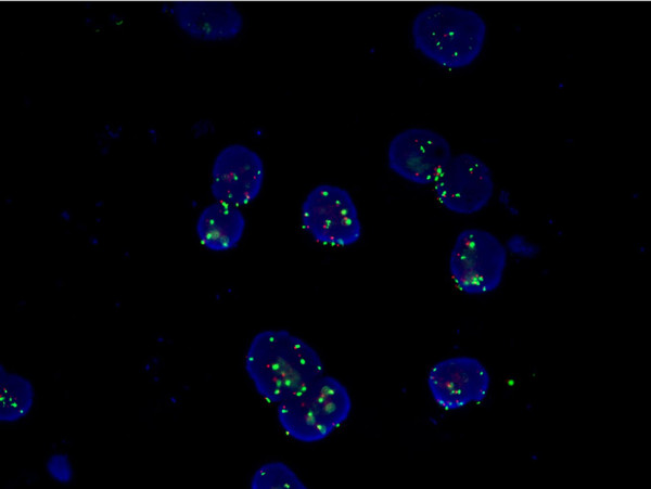
We conducted a retrospective cohort study of 93 inpatients with pleural effusions (72 malignant pleural effusions metastatic from 11 different organs and 21 benign) over 23 months. All the patients came from Chinese northeast areas. Aspirated pleural fluid underwent cytologic examination and fluorescence in situ hybridization (FISH) for aneuploidy. We used FISH in single-colour or if appropriate in dual-colour evaluation to detect chromosomal aberrations (chromosomes 7, 11, and 17) in effusion cells as markers of malignancy, to raise the diagnostic yield and identified the efficiency by diagnostic biopsy. Predominant cytogenetic anomalies and patterns of intratumor cytogenetic heterogeneity were brought in relation to overall survival rate.
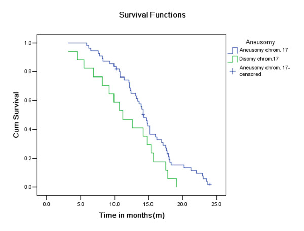
Cytology alone confirmed malignant pleural effusions in 45 of 72 patients (sensitivity 63%), whereas FISH alone positively identified 48 of 72 patients (sensitivity 67%). Both tests had high specificity in predicting benign effusions. If cytology and FISH were considered together, they exhibited 88% sensitivity and 94.5% specificity in discriminating benign and malignant effusions. Combined, the two assays were more sensitive than either test alone. Although the positive predictive value of each test was 94.5%, the negative predictive value of cytology and FISH combined was 78%, better than 47% and 44% for FISH and cytology alone, respectively. There was a significantly prolonged survival rate for patients with aneuploidy for chromosome 17.
FISH in combination with conventional cytology is a highly sensitive and specific diagnostic tool for detecting malignant cells in pleural effusions . The high sensitivity and specificity may be associated with geographic area and race. Simple numeric FISH anomalies may be prognostic.
传统细胞学检查在识别积液中的恶性细胞时,其敏感性和特异性有限,受到了阻碍。本研究的目的是探讨荧光原位杂交(FISH)作为恶性胸腔积液患者传统细胞学检查辅助手段的价值。
我们对93例胸腔积液住院患者进行了一项回顾性队列研究(72例为来自11个不同器官转移的恶性胸腔积液,21例为良性胸腔积液),研究历时23个月。所有患者均来自中国东北地区。抽取的胸腔积液进行了细胞学检查和荧光原位杂交(FISH)以检测非整倍体。我们采用单颜色或适当的双颜色FISH评估来检测积液细胞中的染色体畸变(7号、11号和17号染色体)作为恶性肿瘤的标志物,以提高诊断率,并通过诊断性活检确定其有效性。主要的细胞遗传学异常和肿瘤内细胞遗传学异质性模式与总生存率相关。
仅细胞学检查确诊了72例患者中的45例恶性胸腔积液(敏感性63%),而仅FISH阳性识别出72例患者中的48例(敏感性67%)。两种检查在预测良性积液方面均具有高特异性。如果将细胞学和FISH结合考虑,它们在鉴别良性和恶性积液方面表现出88%的敏感性和94.5%的特异性。两者结合,这两种检测方法比单独任何一种检测都更敏感。虽然每种检测的阳性预测值均为94.5%,但细胞学和FISH联合检测的阴性预测值为78%,分别优于单独FISH的47%和单独细胞学的44%。17号染色体非整倍体患者的生存率显著延长。
FISH与传统细胞学相结合是检测胸腔积液中恶性细胞的一种高度敏感和特异的诊断工具。高敏感性和特异性可能与地理区域和种族有关。简单的数字FISH异常可能具有预后意义。