Department of Cell Biology, Harvard Medical School, Boston, MA 02115, USA.
Mol Cell. 2013 Jan 24;49(2):249-61. doi: 10.1016/j.molcel.2012.11.002. Epub 2012 Dec 6.
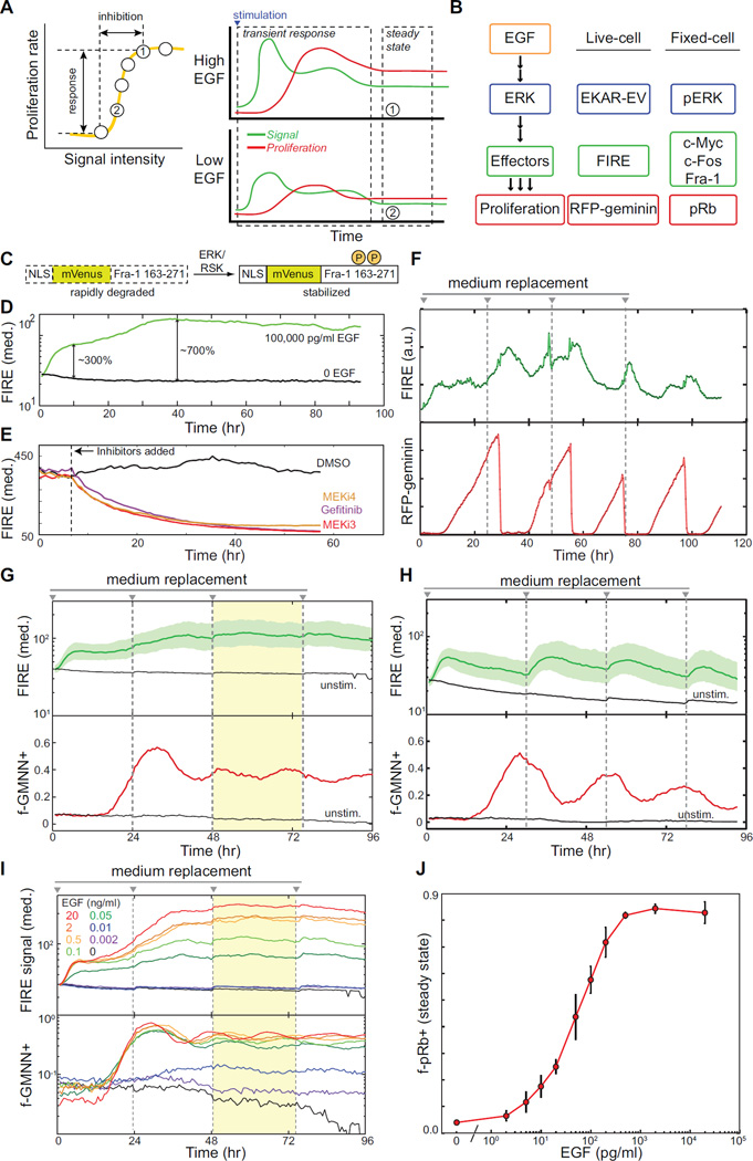
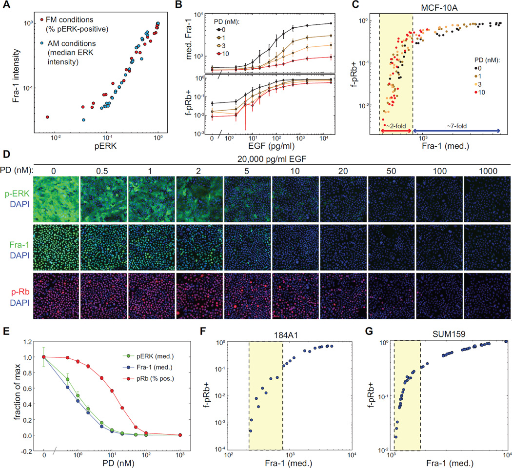
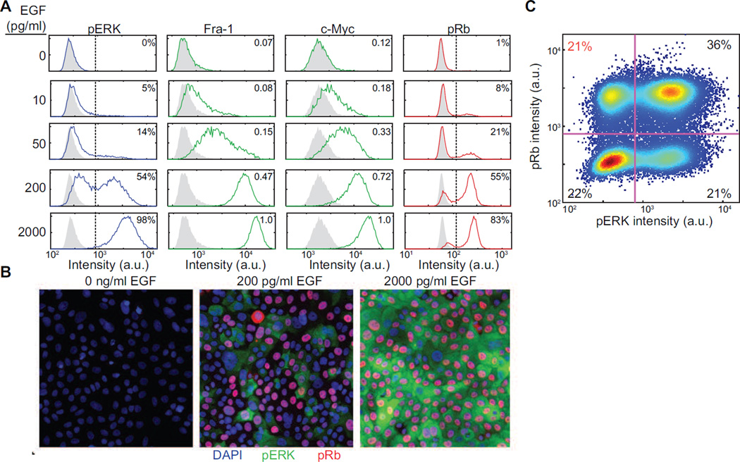
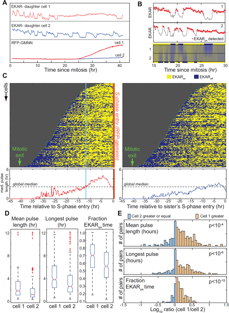
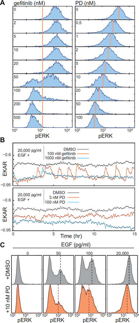
The EGF-stimulated ERK/MAPK pathway is a key conduit for cellular proliferation signals and a therapeutic target in many cancers. Here, we characterize two central quantitative aspects of this pathway: the mechanism by which signal strength is encoded and the response curve relating signal output to proliferation. Under steady-state conditions, we find that ERK is activated in discrete, asynchronous pulses with frequency and duration determined by extracellular concentrations of EGF spanning the physiological range. In genetically identical sister cells, cell-to-cell variability in pulse dynamics influences the decision to enter S phase. While targeted inhibition of EGFR reduces the frequency of ERK activity pulses, inhibition of MEK reduces their amplitude. Continuous response curves measured in multiple cell lines reveal that proliferation is effectively silenced only when ERK pathway output falls below a threshold of ~10%, indicating that high-dose targeting of the pathway is necessary to achieve therapeutic efficacy.
表皮生长因子(EGF)刺激的 ERK/MAPK 通路是细胞增殖信号的关键途径,也是许多癌症的治疗靶点。在这里,我们描述了该通路的两个重要定量方面:信号强度的编码机制以及信号输出与增殖之间的响应曲线。在稳态条件下,我们发现 ERK 以离散、异步的脉冲形式被激活,其频率和持续时间由生理范围内 EGF 的细胞外浓度决定。在遗传上相同的姐妹细胞中,脉冲动力学的细胞间变异性影响进入 S 期的决策。虽然 EGFR 的靶向抑制会降低 ERK 活性脉冲的频率,但 MEK 的抑制会降低其幅度。在多个细胞系中测量的连续响应曲线表明,只有当 ERK 通路输出低于约 10%的阈值时,增殖才会被有效抑制,这表明需要高剂量靶向该通路才能实现治疗效果。