Department of Neurosurgery, Ohio State University, 410 West 10th Avenue, N1025 Doan Hall, Columbus, OH 43210, USA.
J Neurol Neurosurg Psychiatry. 2013 Aug;84(8):843-9. doi: 10.1136/jnnp-2012-303194. Epub 2012 Dec 15.
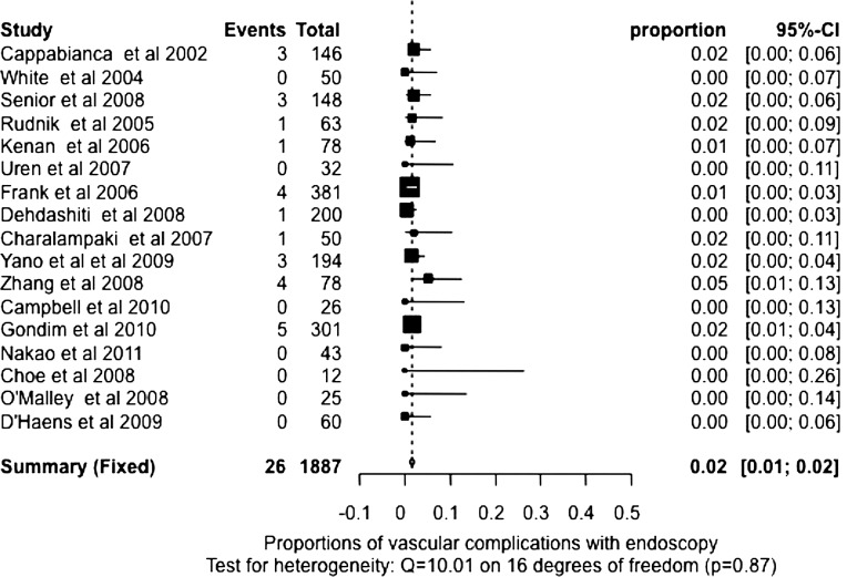
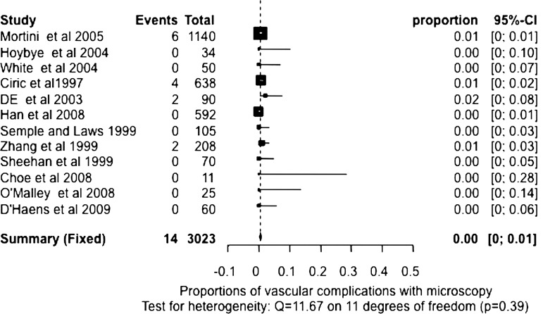
Endoscopic transsphenoidal pituitary surgery has become increasingly more popular for the removal of pituitary adenomas. It is also widely recognised that transsphenoidal microscopic removal of pituitary adenomas is a well-established procedure with good outcomes. Our objective was to meta-analyse the short-term results of endoscopic and microscopic pituitary adenoma surgery. We undertook a systematic review of the English literature on results of transsphenoidal surgery, both microscopic and endoscopic from 1990 to 2011. Series with less than 10 patients were excluded. Pooled data were analysed using meta-analysis techniques to obtain estimate of death, complication rates and extent of tumour removal. Complications evaluated included cerebrospinal fluid leak, meningitis, vascular complications, visual complications, diabetes insipidus, hypopituitarism and cranial nerve injury. Data were also analysed for tumour size and sex. 38 studies met the inclusion criteria yielding 24 endoscopic and 22 microscopic datasets (eight studies included both endoscopic and microscopic series). Meta-analysis of the available literature showed that the endoscopic transsphenoidal technique was associated with a higher incidence of vascular complications (p<0.0001). No difference was found between the two techniques in all other variables examined. Meta-analysis of the available literature reveals that endoscopic removal of pituitary adenoma, in the short term, does not seem to confer any advantages over the microscopic technique and the incidence of reported vascular complications was higher with endoscopic than with microscopic removal of pituitary adenomas. While we recognise the limitations of meta-analysis, our study suggests that a multicentre, randomised, comparative effectiveness study of the microscopic and endoscopic transsphenoidal techniques may be a reasonable approach towards establishing a true valuation of these techniques.
经蝶窦垂体手术已越来越多地用于切除垂体腺瘤。广泛认为,经蝶窦显微镜下切除垂体腺瘤是一种成熟的手术方法,效果良好。我们的目的是对内镜和显微镜下垂体腺瘤手术的短期结果进行荟萃分析。我们对 1990 年至 2011 年间经蝶窦手术(显微镜和内镜)结果的英文文献进行了系统评价,排除了少于 10 例患者的系列研究。使用荟萃分析技术对汇总数据进行分析,以获得死亡、并发症发生率和肿瘤切除程度的估计值。评估的并发症包括脑脊液漏、脑膜炎、血管并发症、视觉并发症、尿崩症、垂体功能减退和颅神经损伤。数据还按肿瘤大小和性别进行了分析。38 项研究符合纳入标准,其中包括 24 项内镜和 22 项显微镜数据集(8 项研究包括内镜和显微镜系列)。对现有文献的荟萃分析表明,内镜经蝶窦技术与更高的血管并发症发生率相关(p<0.0001)。在所有其他检查变量中,两种技术之间没有差异。对现有文献的荟萃分析表明,短期内内镜切除垂体腺瘤似乎并没有比显微镜技术带来任何优势,而且内镜切除垂体腺瘤的血管并发症发生率高于显微镜切除。虽然我们认识到荟萃分析的局限性,但我们的研究表明,对显微镜和内镜经蝶窦技术进行多中心、随机、对照有效性研究可能是一种合理的方法,可以对这些技术进行真正的评估。