• 文献检索
  • 文档翻译
  • 深度研究
  • 学术资讯
  • Suppr Zotero 插件Zotero 插件
  • 邀请有礼
  • 套餐&价格
  • 历史记录
应用&插件
Suppr Zotero 插件Zotero 插件浏览器插件Mac 客户端Windows 客户端微信小程序
定价
高级版会员购买积分包购买API积分包
服务
文献检索文档翻译深度研究API 文档MCP 服务
关于我们
关于 Suppr公司介绍联系我们用户协议隐私条款
关注我们

Suppr 超能文献

核心技术专利:CN118964589B侵权必究
粤ICP备2023148730 号-1Suppr @ 2026

文献检索

告别复杂PubMed语法,用中文像聊天一样搜索,搜遍4000万医学文献。AI智能推荐,让科研检索更轻松。

立即免费搜索

文件翻译

保留排版,准确专业,支持PDF/Word/PPT等文件格式,支持 12+语言互译。

免费翻译文档

深度研究

AI帮你快速写综述,25分钟生成高质量综述,智能提取关键信息,辅助科研写作。

立即免费体验

全身 MRI 和锝-99m 亚甲基二膦酸盐骨扫描评估多发性骨髓瘤疾病进展和预后:一项前瞻性对比研究。

Assessment of whole body MRI and sestamibi technetium-99m bone marrow scan in prediction of multiple myeloma disease progression and outcome: a prospective comparative study.

机构信息

Department of Medicine and Clinical Haematology, Launceston General Hospital, Launceston, Tasmania, Australia.

出版信息

BMJ Open. 2013 Jan 10;3(1):e002025. doi: 10.1136/bmjopen-2012-002025.

DOI:10.1136/bmjopen-2012-002025
PMID:23315438
原文链接:https://pmc.ncbi.nlm.nih.gov/articles/PMC3549203/
Abstract

OBJECTIVES

This study aims primarily to determine whether whole body MRI (WB-MRI) and Sestamibi Technetium-99m-bone marrow (MIBI) scans in the same patients produce the same estimate of disease load and location, and secondly, to study possible association between the bone disease detected by these scans and the effect on disease outcome and survival. Bone disease occurs in about 90% of multiple myeloma (MM) patients. There are no data comparing the new diagnostic modalities with WB-MRI and MIBI in MM.

DESIGN

A prospective comparative study between WB-MRI and MIBI scans in assessing bone disease and outcome of MM.

PARTICIPANTS AND METHODS

Sixty-two consecutive patients with confirmed MM underwent simultaneous WB-MRI (both axial T1 and turbo spin echo short tau inversion recovery (STIR)) and MIBI scans at a single institution from January 2010 to January 2011, and their survival status was determined in January 2012. The median age was 62 years (range 37-88) with a male-to-female ratio of 33 : 29.

RESULTS

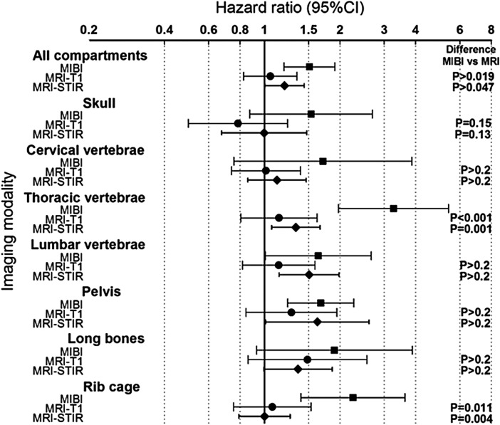
In vertebrae and long bones, MRI scan detected more disease compared with MIBI scan (p<0.001) but there was less difference in the skull (p=0.09). In the ribcage, the MIBI scan detected more lytic lesions of the ribs compared with MRI scan (p<0.001). Thirteen of the 62 patients died during the 24-month follow-up. Increased disease detected in all bones by both scans was associated with increased mortality risk (MIBI p=0.001; MRI-STIR p=0.044; but not MRI-T1 p=0.44). In all combined bone groups, the mean MIBI scan results provided a better prediction of mortality than MRI scan over the follow-up period (MRI-T1 vs MIBI p=0.019; MRI-STIR vs MIBI p=0.047).

CONCLUSIONS

Although WB-MRI detected more MM bone disease, MIBI scan predicted overall disease outcome and mortality better than MRI scan. Further studies to define optimum use of these imaging techniques are warranted.

TRIAL REGISTRATION NUMBER

The study was registered prospectively in the Australian and New Zealand Clinical Trials Registry at http://www.ANZCTR.org.au under No: ACTRN12609000761268.

摘要

目的

本研究旨在首先确定同一患者的全身磁共振成像(WB-MRI)和锝 99m 亚甲基二膦酸盐 Sestamibi 扫描是否能产生相同的疾病负荷和位置估计,其次,研究这些扫描发现的骨疾病与疾病结果和生存之间的可能关联。多发性骨髓瘤(MM)患者约有 90%发生骨病。目前尚无比较这些新诊断方法与 MM 中 WB-MRI 和 MIBI 的资料。

设计

在单一机构内,于 2010 年 1 月至 2011 年 1 月,对 62 例确诊的 MM 患者同时进行了 WB-MRI(轴位 T1 和涡轮自旋回波短 tau 反转恢复(STIR))和 MIBI 扫描,2012 年 1 月确定了他们的生存状况。中位年龄为 62 岁(范围 37-88 岁),男女比例为 33︰29。

结果

在椎体和长骨中,MRI 扫描比 MIBI 扫描检测到更多的病变(p<0.001),但颅骨的差异较小(p=0.09)。在肋骨中,MIBI 扫描比 MRI 扫描检测到更多的肋骨溶骨性病变(p<0.001)。在 24 个月的随访期间,62 例患者中有 13 例死亡。两种扫描均在所有骨骼中检测到更多的疾病与死亡率升高相关(MIBI,p=0.001;MRI-STIR,p=0.044;但 MRI-T1,p=0.44 未显示差异)。在所有综合骨骼组中,在整个随访期间,MIBI 扫描的平均结果比 MRI 扫描更能预测死亡率(MRI-T1 与 MIBI,p=0.019;MRI-STIR 与 MIBI,p=0.047)。

结论

尽管 WB-MRI 检测到更多的 MM 骨疾病,但 MIBI 扫描比 MRI 扫描更能预测总体疾病结局和死亡率。需要进一步的研究来确定这些影像学技术的最佳应用。

试验注册

该研究在澳大利亚和新西兰临床试验注册中心(http://www.ANZCTR.org.au)进行了前瞻性注册,编号为 ACTRN12609000761268。

https://cdn.ncbi.nlm.nih.gov/pmc/blobs/e031/3549203/b1424f775dec/bmjopen2012002025f03.jpg
https://cdn.ncbi.nlm.nih.gov/pmc/blobs/e031/3549203/01adab6f2ade/bmjopen2012002025f01.jpg
https://cdn.ncbi.nlm.nih.gov/pmc/blobs/e031/3549203/9b88b89112ea/bmjopen2012002025f02.jpg
https://cdn.ncbi.nlm.nih.gov/pmc/blobs/e031/3549203/b1424f775dec/bmjopen2012002025f03.jpg
https://cdn.ncbi.nlm.nih.gov/pmc/blobs/e031/3549203/01adab6f2ade/bmjopen2012002025f01.jpg
https://cdn.ncbi.nlm.nih.gov/pmc/blobs/e031/3549203/9b88b89112ea/bmjopen2012002025f02.jpg
https://cdn.ncbi.nlm.nih.gov/pmc/blobs/e031/3549203/b1424f775dec/bmjopen2012002025f03.jpg

相似文献

1
Assessment of whole body MRI and sestamibi technetium-99m bone marrow scan in prediction of multiple myeloma disease progression and outcome: a prospective comparative study.全身 MRI 和锝-99m 亚甲基二膦酸盐骨扫描评估多发性骨髓瘤疾病进展和预后:一项前瞻性对比研究。
BMJ Open. 2013 Jan 10;3(1):e002025. doi: 10.1136/bmjopen-2012-002025.
2
Technetium-99m 2-methoxy-isobutyl-isonitrile uptake scintigraphy in detection of the bone marrow infiltration in multiple myeloma: correlation with MRI and other prognostic factors.锝-99m甲氧基异丁基异腈摄取闪烁扫描术在检测多发性骨髓瘤骨髓浸润中的应用:与MRI及其他预后因素的相关性
Ann Hematol. 2007 Nov;86(11):805-13. doi: 10.1007/s00277-007-0329-z. Epub 2007 Aug 8.
3
Multiple myeloma: predictive value of Tc-99m MIBI scintigraphy and MRI in its diagnosis and therapy.多发性骨髓瘤:锝-99m甲氧基异丁基异腈闪烁扫描术和磁共振成像在其诊断与治疗中的预测价值
Nucl Med Rev Cent East Eur. 2008;11(1):12-6.
4
Value of Tc-99m sestamibi scintigraphy in the detection of bone lesions in multiple myeloma: comparison with Tc-99m methylene diphosphonate.锝-99m 甲氧基异丁基异腈闪烁显像在检测多发性骨髓瘤骨病变中的价值:与锝-99m 亚甲基二膦酸盐的比较
Ann Hematol. 2001 Jun;80(6):349-53. doi: 10.1007/s002770100302.
5
18F-FDG PET/CT, 99mTc-MIBI, and MRI in evaluation of patients with multiple myeloma.18F-FDG PET/CT、99mTc-MIBI及MRI在多发性骨髓瘤患者评估中的应用
J Nucl Med. 2008 Feb;49(2):195-200. doi: 10.2967/jnumed.107.045641. Epub 2008 Jan 16.
6
Technetium-99m sestamibi scintigraphy is sensitive and specific for the staging and the follow-up of patients with multiple myeloma: a multicentre study on 397 scans.锝-99m 甲氧基异丁基异腈闪烁扫描术对多发性骨髓瘤患者的分期及随访具有敏感性和特异性:一项对397次扫描的多中心研究。
Br J Haematol. 2007 Mar;136(5):729-35. doi: 10.1111/j.1365-2141.2006.06489.x.
7
Technetium-99m-sestamibi scintigraphy: an alternative approach for diagnosis and follow-up of active myeloma lesions after high-dose chemotherapy and autologous stem cell transplantation.锝-99m-甲氧基异丁基异腈闪烁扫描术:一种用于大剂量化疗和自体干细胞移植后活动性骨髓瘤病灶诊断及随访的替代方法。
Ann Hematol. 2001 Jul;80(7):393-7. doi: 10.1007/s002770100318.
8
A comparison of fluorine-18 fluoro-deoxyglucose PET and technetium-99m sestamibi in assessing patients with multiple myeloma.氟-18氟脱氧葡萄糖正电子发射断层扫描(PET)与锝-99m甲氧基异丁基异腈在评估多发性骨髓瘤患者中的比较。
Eur J Haematol. 2004 Jan;72(1):32-7. doi: 10.1046/j.0902-4441.2004.00177.x.
9
18F-FDG PET/CT, 99mTc-MIBI, and MRI in the prediction of outcome of patients with multiple myeloma: a comparative study.18F-FDG PET/CT、99mTc-MIBI 和 MRI 对多发性骨髓瘤患者预后的预测作用:一项比较研究。
Clin Nucl Med. 2015 Apr;40(4):303-8. doi: 10.1097/RLU.0000000000000696.
10
99mTc-sestamibi imaging and bone marrow karyotyping in the assessment of multiple myeloma and MGUS.99mTc-司他米比显像和骨髓核型分析在多发性骨髓瘤和意义未明的单克隆丙种球蛋白血症评估中的应用
Nucl Med Commun. 2008 Jun;29(6):535-41. doi: 10.1097/MNM.0b013e3282f5e5df.

引用本文的文献

1
99mTc-sestamibi SPECT/CT and 18F-FDG-PET/CT have similar performance but different imaging patterns in newly diagnosed multiple myeloma.99mTc- 美罗昔单抗 SPECT/CT 和 18F-FDG-PET/CT 在新诊断的多发性骨髓瘤中的表现相似,但成像模式不同。
Nucl Med Commun. 2020 Oct;41(10):1081-1088. doi: 10.1097/MNM.0000000000001259.

本文引用的文献

1
Imaging in multiple myeloma.多发性骨髓瘤的影像学检查
Recent Results Cancer Res. 2011;183:133-47. doi: 10.1007/978-3-540-85772-3_7.
2
Current concepts in the evaluation of multiple myeloma with MR imaging and FDG PET/CT.MR 成像和 FDG PET/CT 评估多发性骨髓瘤的当前概念。
Radiographics. 2010 Jan;30(1):127-42. doi: 10.1148/rg.301095066.
3
Role of radiography, MRI and FDG-PET/CT in diagnosing, staging and therapeutical evaluation of patients with multiple myeloma.X线摄影、磁共振成像及氟代脱氧葡萄糖正电子发射断层显像/计算机断层扫描在多发性骨髓瘤患者诊断、分期及治疗评估中的作用
Ann Hematol. 2009 Dec;88(12):1161-8. doi: 10.1007/s00277-009-0829-0.
4
International myeloma working group consensus statement and guidelines regarding the current role of imaging techniques in the diagnosis and monitoring of multiple Myeloma.国际骨髓瘤工作组关于成像技术在多发性骨髓瘤诊断和监测中当前作用的共识声明及指南。
Leukemia. 2009 Sep;23(9):1545-56. doi: 10.1038/leu.2009.89. Epub 2009 May 7.
5
Skeletal imaging and management of bone disease.骨骼成像与骨病管理
Hematology Am Soc Hematol Educ Program. 2008:313-9. doi: 10.1182/asheducation-2008.1.313.
6
Pathogenesis of myeloma bone disease.骨髓瘤骨病的发病机制。
Leukemia. 2009 Mar;23(3):435-41. doi: 10.1038/leu.2008.336. Epub 2008 Nov 27.
7
Whole-body MRI versus whole-body MDCT for staging of multiple myeloma.全身磁共振成像与全身多层螺旋计算机断层扫描在多发性骨髓瘤分期中的应用比较
AJR Am J Roentgenol. 2008 Apr;190(4):1097-104. doi: 10.2214/AJR.07.2635.
8
18F-FDG PET/CT, 99mTc-MIBI, and MRI in evaluation of patients with multiple myeloma.18F-FDG PET/CT、99mTc-MIBI及MRI在多发性骨髓瘤患者评估中的应用
J Nucl Med. 2008 Feb;49(2):195-200. doi: 10.2967/jnumed.107.045641. Epub 2008 Jan 16.
9
Guidelines for the use of imaging in the management of myeloma.骨髓瘤管理中影像学检查应用指南。
Br J Haematol. 2007 Apr;137(1):49-63. doi: 10.1111/j.1365-2141.2007.06491.x.
10
Technetium-99m sestamibi scintigraphy is sensitive and specific for the staging and the follow-up of patients with multiple myeloma: a multicentre study on 397 scans.锝-99m 甲氧基异丁基异腈闪烁扫描术对多发性骨髓瘤患者的分期及随访具有敏感性和特异性:一项对397次扫描的多中心研究。
Br J Haematol. 2007 Mar;136(5):729-35. doi: 10.1111/j.1365-2141.2006.06489.x.