VCU Pauley Heart Center, Virginia Commonwealth University, Richmond, VA 23298, USA.
J Am Heart Assoc. 2012 Oct;1(5):e002360. doi: 10.1161/JAHA.112.002360. Epub 2012 Oct 25.
Despite the clear advantages of reperfusion in acute myocardial infarction, part of the myocardium is injured during reperfusion by reactive oxygen species. Reactive oxygen species activate apoptosis signal-regulating kinase-1, a key mediator in cell death. We hypothesized that inhibition of apoptosis signal-regulating kinase-1 at the time of reperfusion would protect the heart from ischemia-reperfusion injury.
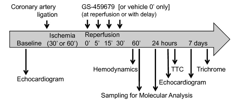
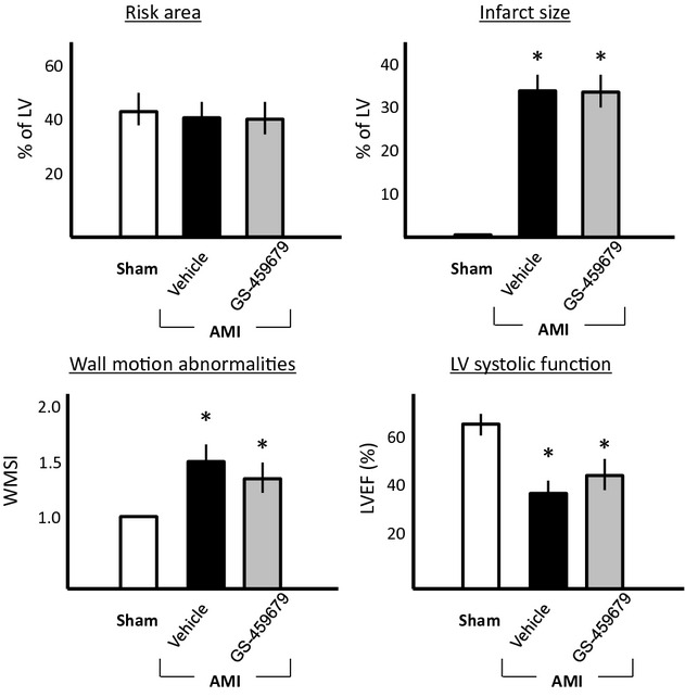
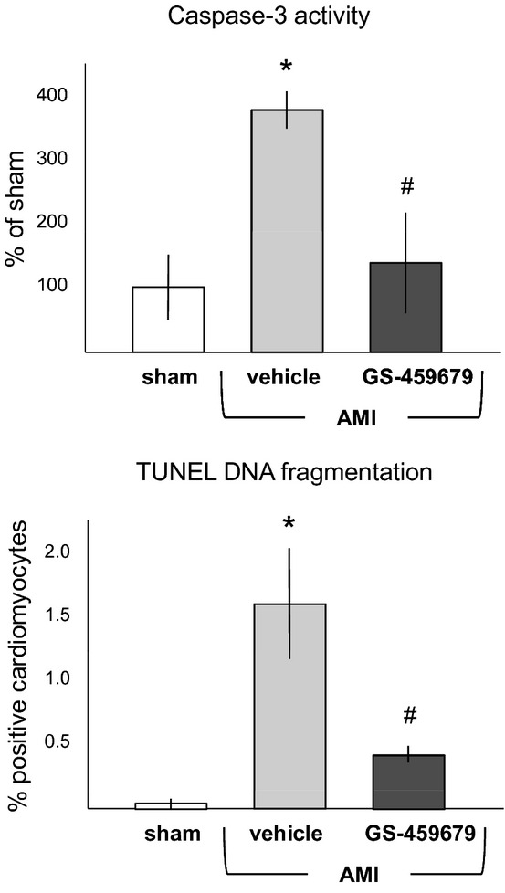
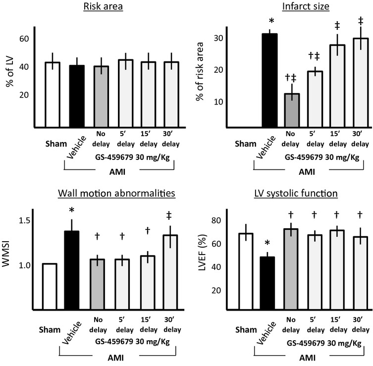
Male CD1 mice underwent transient coronary artery ligation (30 minutes) followed by reperfusion or underwent sham surgery (n=10 to 12 per group). A selective small-molecule inhibitor of apoptosis signal-regulating kinase-1 (GS-459679) was given immediately after reperfusion (10 or 30 mg/kg IP). Infarct size was measured early (at 24 hours, in a subgroup of mice) by triphenyl tetrazolium chloride staining and late (at 7 days) by Masson's trichrome staining for fibrosis. Apoptosis was assessed by measurement of caspase-3 activity and by determination of DNA fragmentation in cardiomyocytes bordering the infarct. Transthoracic echocardiography was performed before surgery and then at 24 hours and 7 days later. Treatment with GS-459679 at reperfusion led to a significant dose-related reduction in infarct size (31% for 10 mg/kg [P<0.001 versus vehicle] and 60% for 30 mg/kg [P<0.001 versus vehicle]), inhibition of apoptotic cell death, and preservation of left ventricular dimension and systolic function at both 24 hours and 7 days.
Inhibition of apoptosis signal-regulating kinase-1 at the time of reperfusion limits infarct size and preserves left ventricular function in a model of acute myocardial infarction in the mouse.
尽管在急性心肌梗死中再灌注具有明显的优势,但部分心肌在再灌注过程中会受到活性氧的损伤。活性氧会激活细胞死亡的关键介质凋亡信号调节激酶-1。我们假设在再灌注时抑制凋亡信号调节激酶-1会保护心脏免受缺血再灌注损伤。
雄性 CD1 小鼠接受短暂的冠状动脉结扎(30 分钟),然后进行再灌注或接受假手术(每组 10 至 12 只)。在再灌注后立即给予凋亡信号调节激酶-1 的选择性小分子抑制剂(GS-459679,10 或 30mg/kg,腹腔内注射)。通过三苯基四氮唑氯化物染色在早期(在亚组小鼠中 24 小时)测量梗死面积,并用 Masson 三色染色在晚期(7 天)测量纤维化。通过测定半胱天冬酶-3 活性和测定梗死边缘的心肌细胞 DNA 片段来评估凋亡。在手术前、24 小时和 7 天后进行经胸超声心动图检查。再灌注时给予 GS-459679 治疗可显著剂量依赖性地减少梗死面积(10mg/kg 时为 31%[P<0.001 与载体相比],30mg/kg 时为 60%[P<0.001 与载体相比]),抑制细胞凋亡死亡,并在 24 小时和 7 天后保留左心室维度和收缩功能。
在急性心肌梗死模型中,再灌注时抑制凋亡信号调节激酶-1可限制梗死面积并保护左心室功能。