Department of Biochemistry, Molecular Biology and Biophysics, University of Minnesota, Minneapolis, MN 55455, USA.
J Mol Biol. 2013 Apr 12;425(7):1172-82. doi: 10.1016/j.jmb.2013.01.010. Epub 2013 Jan 11.
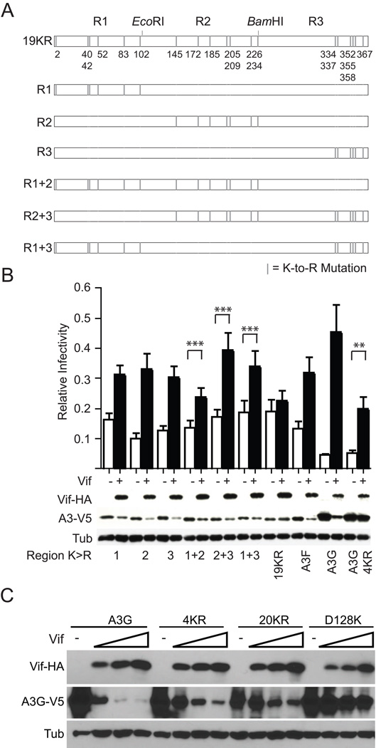
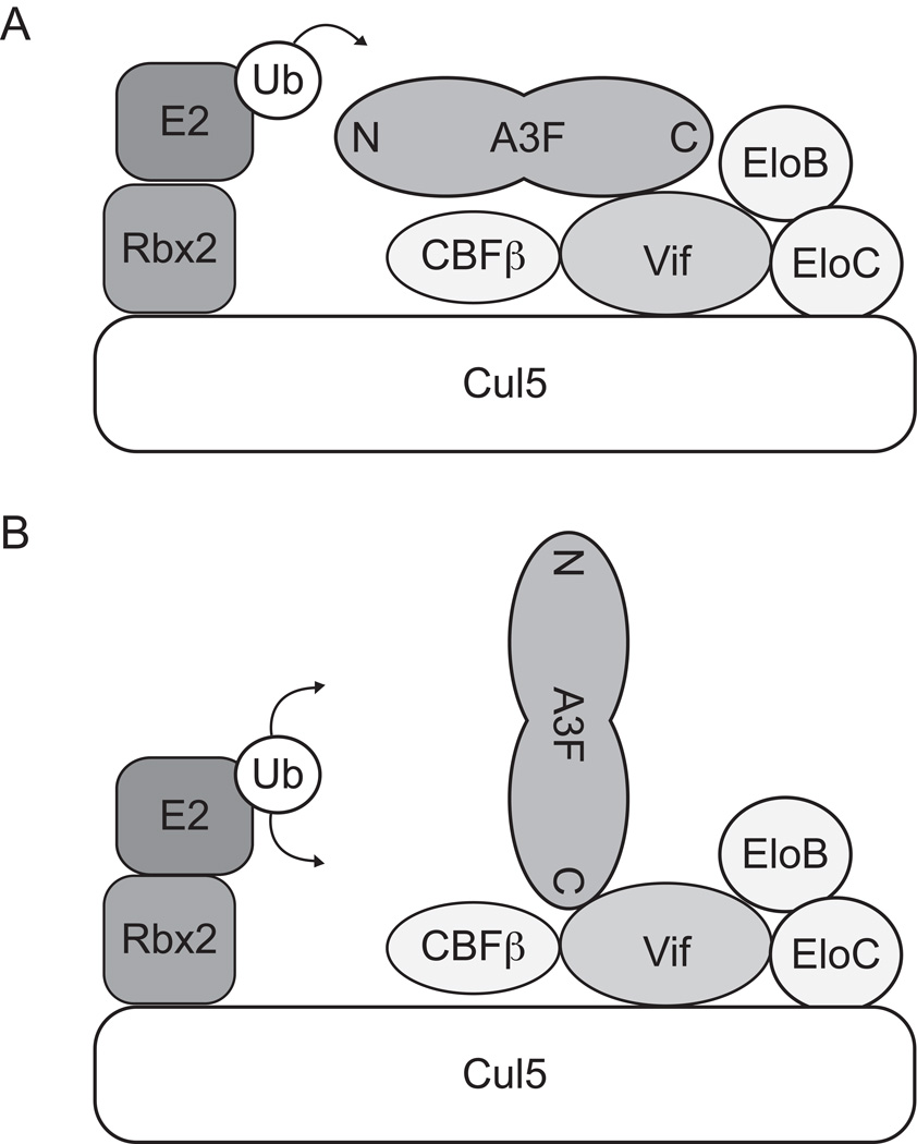
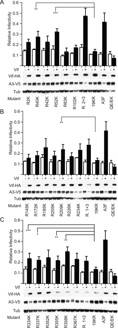
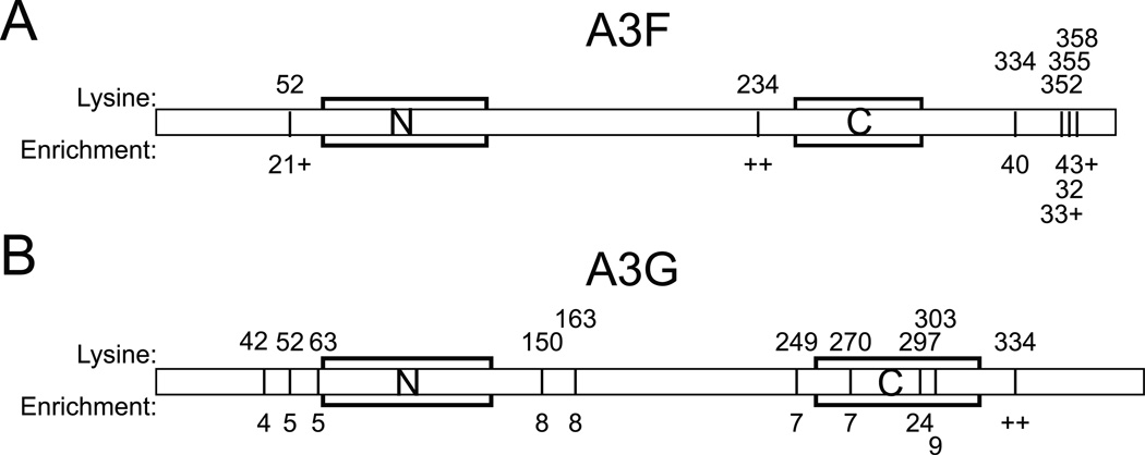
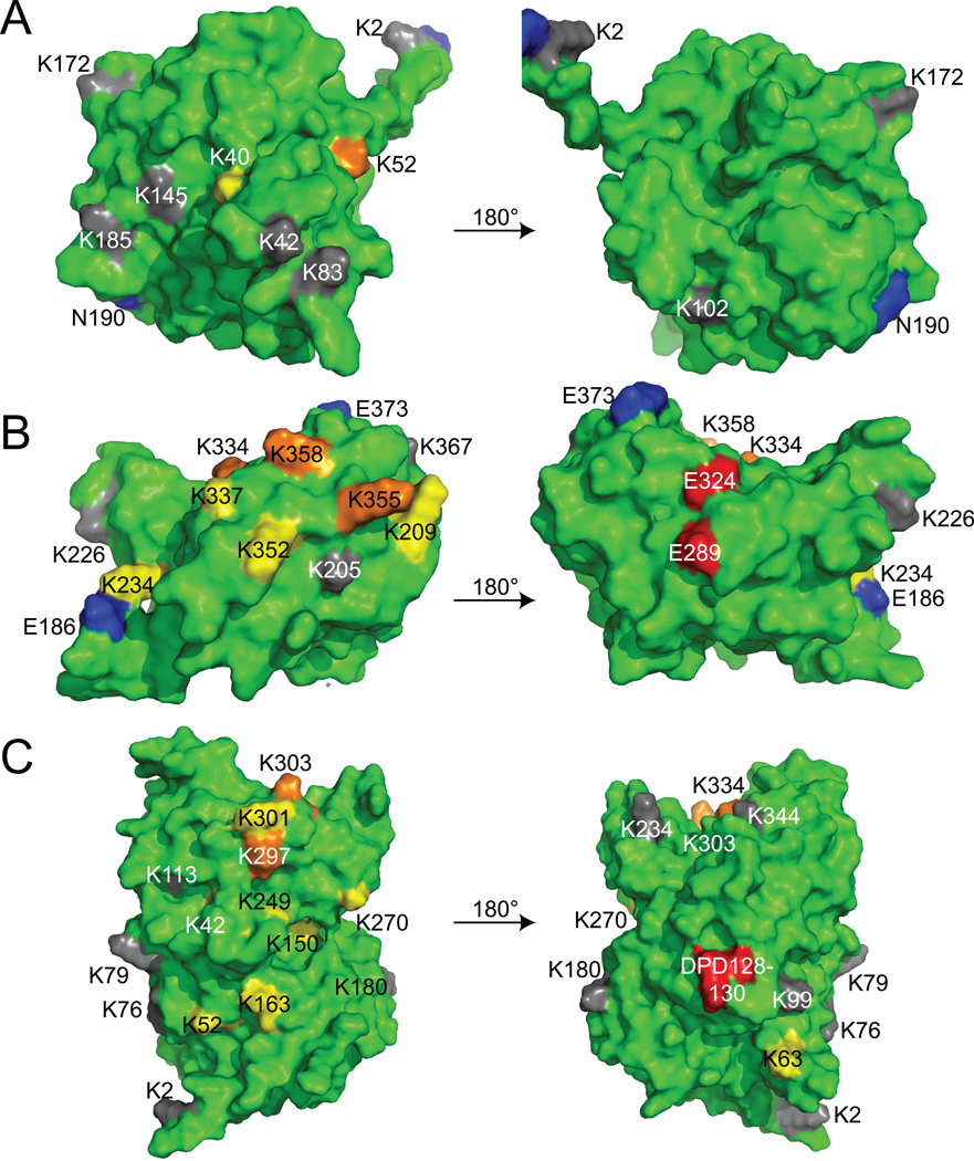
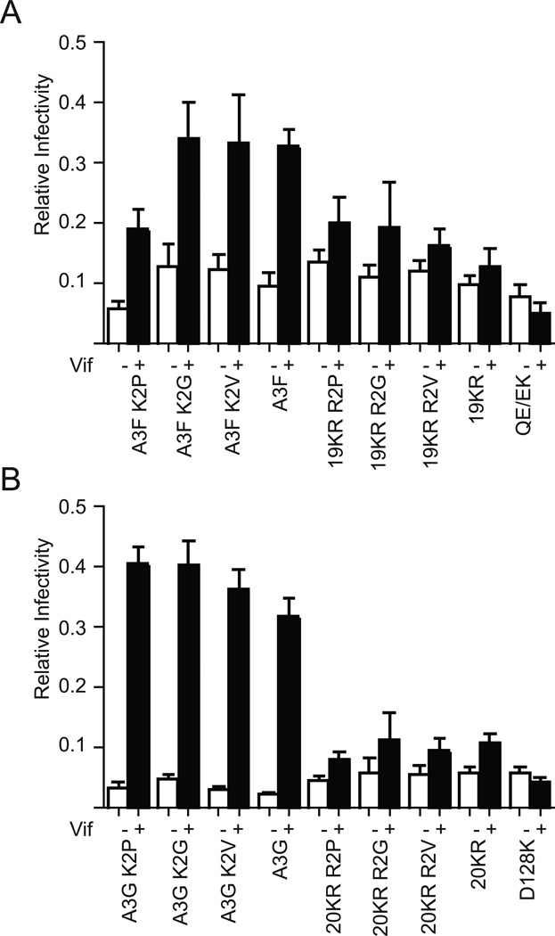
APOBEC3F (A3F) and APOBEC3G (A3G) are DNA cytosine deaminases that potently restrict human immunodeficiency virus type 1 replication when the virus is deprived of its accessory protein Vif (virion infectivity factor). Vif counteracts these restriction factors by recruiting A3F and A3G to an E3 ubiquitin (Ub) ligase complex that mediates their polyubiquitination (polyUb) and proteasomal degradation. While previous efforts have identified single amino acid residues in APOBEC3 proteins required for Vif recognition, less is known about the downstream Ub acceptor sites that are targeted. One prior report identified a cluster of polyubiquitinated residues in A3G and proposed an antiparallel model of A3G interaction with the Vif-E3 Ub ligase complex wherein Vif binding at one terminus of A3G orients the opposite terminus for polyUb [Iwatani et al. (2009). Proc. Natl. Acad. Sci. USA, 106, 19539-19544]. To test the generalizability of this model, we carried out a complete mutagenesis of the lysine residues in A3F and used a complementary, unbiased proteomic approach to identify Ub acceptor sites targeted by Vif. Our data indicate that internal lysines are the dominant Ub acceptor sites in both A3F and A3G. In contrast with the proposed antiparallel model, however, we find that the Vif-dependent polyUb of A3F and A3G can occur at multiple acceptor sites dispersed along predicted lysine-enriched surfaces of both the N- and C-terminal deaminase domains. These data suggest an alternative model for binding of APOBEC3 proteins to the Vif-E3 Ub ligase complex and diminish enthusiasm for the amenability of APOBEC3 Ub acceptor sites to therapeutic intervention.
APOBEC3F(A3F)和 APOBEC3G(A3G)是 DNA 胞嘧啶脱氨酶,当病毒缺乏辅助蛋白 Vif(病毒感染因子)时,它们能有效地限制人类免疫缺陷病毒 1 型的复制。Vif 通过招募 A3F 和 A3G 到一个 E3 泛素(Ub)连接酶复合物来抵消这些限制因子,该复合物介导它们的多泛素化(polyUb)和蛋白酶体降解。虽然以前的研究已经确定了 APOBEC3 蛋白中用于 Vif 识别的单个氨基酸残基,但对靶向的下游 Ub 接受位点知之甚少。之前有一份报告鉴定了 A3G 中多泛素化残基的簇,并提出了 A3G 与 Vif-E3 Ub 连接酶复合物相互作用的反平行模型,其中 Vif 在 A3G 一端的结合使另一端定向进行多泛素化[Iwatani 等人。(2009 年)。Proc。Natl。Acad。Sci。美国,106,19539-19544]。为了测试该模型的普遍性,我们对 A3F 中的赖氨酸残基进行了完全突变,并使用互补的、无偏的蛋白质组学方法来鉴定被 Vif 靶向的 Ub 接受位点。我们的数据表明,内部赖氨酸是 A3F 和 A3G 中主要的 Ub 接受位点。然而,与提出的反平行模型相反,我们发现 A3F 和 A3G 的 Vif 依赖性多泛素化可以发生在分散在两个 N-和 C-末端脱氨酶结构域富含赖氨酸表面上的多个接受位点上。这些数据表明了 APOBEC3 蛋白与 Vif-E3 Ub 连接酶复合物结合的替代模型,并降低了对 APOBEC3 Ub 接受位点进行治疗干预的可行性的热情。