Saati Samira, Nikkerdar Nafiseh, Golshah Amin
Department of Oral and Maxillofacial Radiology, School of Dentistry, Boali University of Medical Sciences, Hamedan, Iran.
Iran J Radiol. 2011 Dec;8(4):253-7. doi: 10.5812/iranjradiol.4588. Epub 2011 Dec 25.
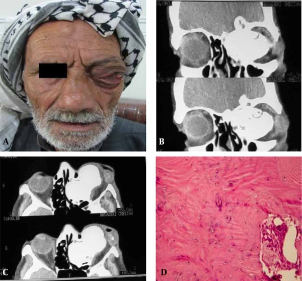
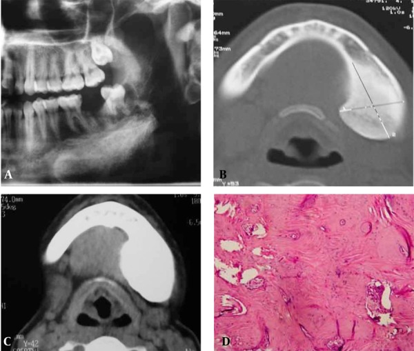
Osteomas are benign osteogenic neoplasms or hamartomas with a very slow growth rate. Osteoma is the most common mesenchymal neoplasm of the paranasal sinuses. In the jaws, the mandible is more commonly involved than the maxilla. Osteomas may occur at any age, but most frequently are found in individuals older than 40 years. Although most osteomas are small, some may become large enough to cause severe damage, especially those that develop in the frontoethmoid region. Osteomas composed solely of compact bone are uniformly radiopaque and those containing cancellous bone show evidence of internal trabecular structure. To determine and evaluate the exact extension and internal structure of these lesions, computed tomography (CT) is a more useful imaging modality in comparison to conventional radiography. Hereby, we discuss clinical and imaging features of two osteomas (one in the ethmoid sinus and the other in the mandible) along with the main differential diagnoses and pathologic features.
骨瘤是良性成骨性肿瘤或错构瘤,生长速度非常缓慢。骨瘤是鼻窦最常见的间叶性肿瘤。在颌骨中,下颌骨比上颌骨更常受累。骨瘤可发生于任何年龄,但最常见于40岁以上的人群。虽然大多数骨瘤较小,但有些可能会长得足够大,造成严重损害,尤其是那些发生在额筛区域的骨瘤。仅由密质骨组成的骨瘤在X线片上表现为均匀的不透光,而含有松质骨的骨瘤则显示出内部小梁结构的迹象。与传统X线摄影相比,计算机断层扫描(CT)在确定和评估这些病变的确切范围和内部结构方面是一种更有用的成像方式。在此,我们讨论两例骨瘤(一例位于筛窦,另一例位于下颌骨)的临床和影像学特征,以及主要的鉴别诊断和病理特征。